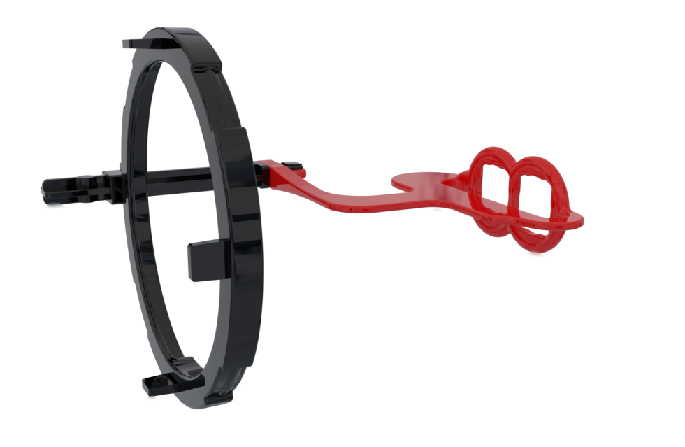
Bitewing
For molar and canine bitewing images.

Our Technology
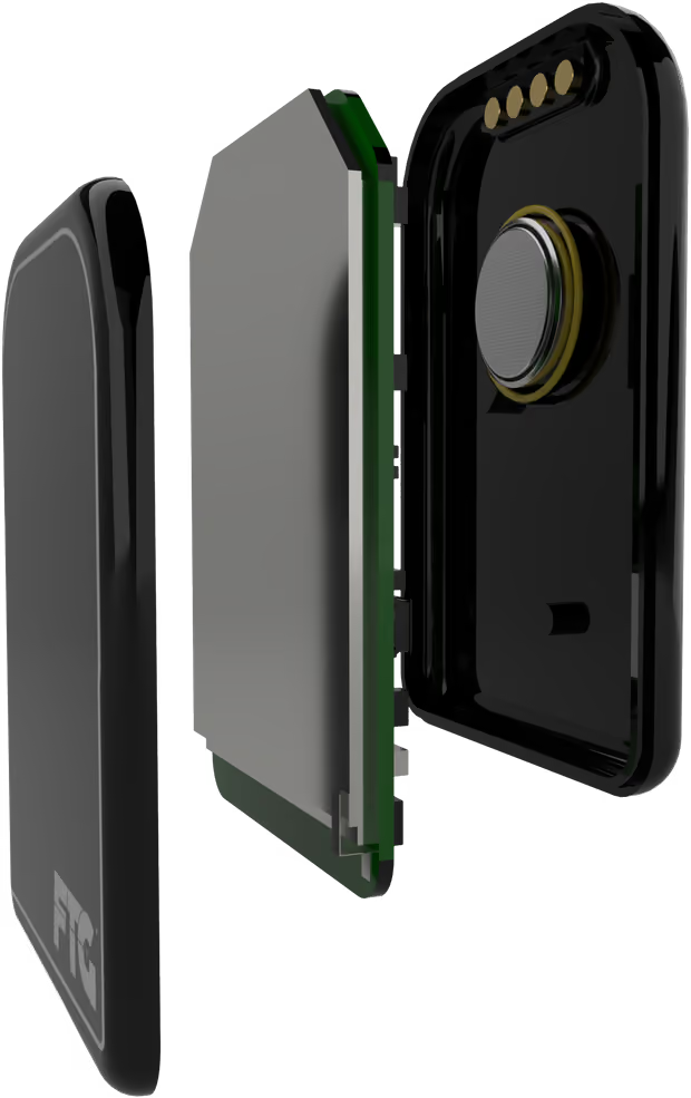
DC-Air® brings revolutionary direct-conversion and wireless technology to dental X-ray sensors.
Request information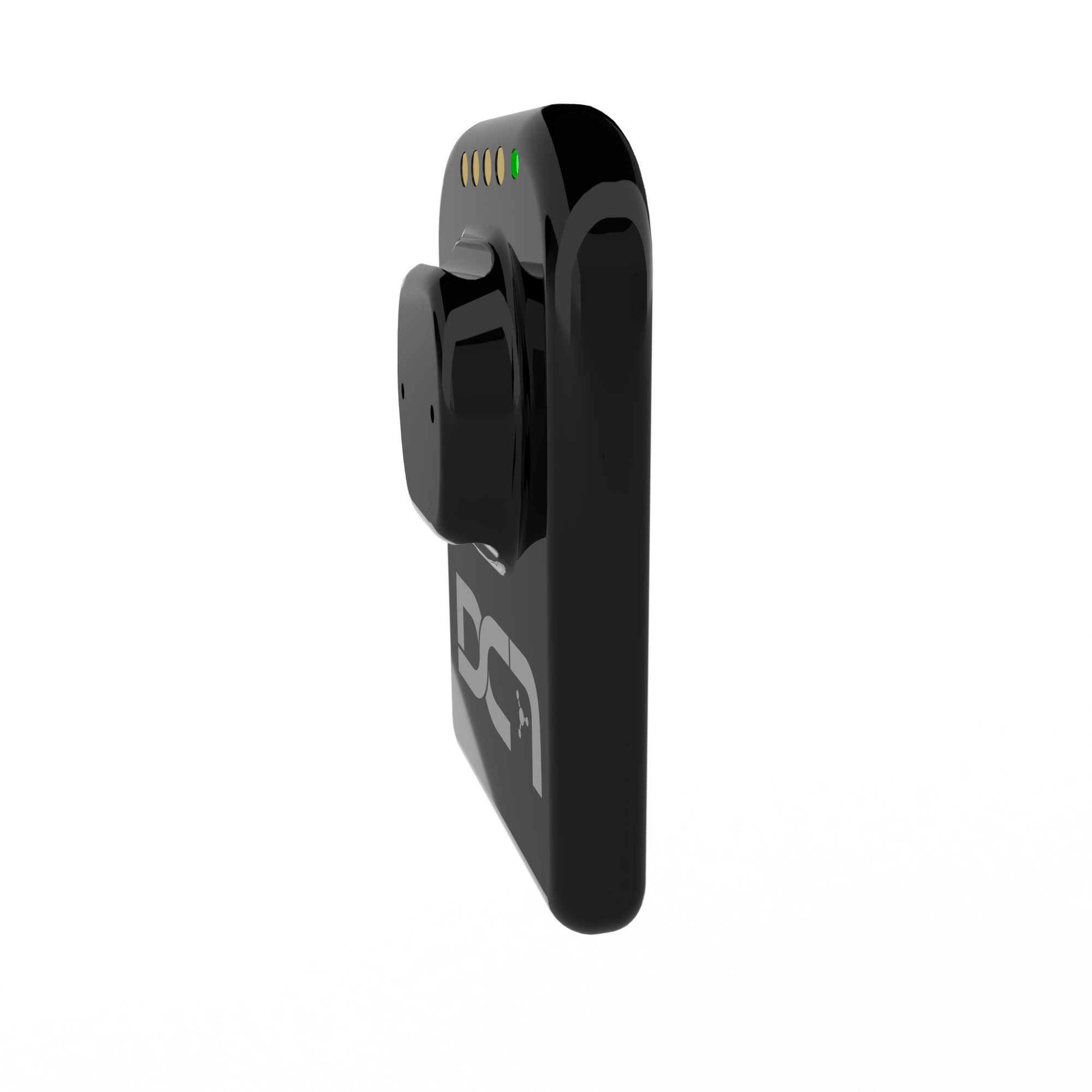
True Wireless® imaging
DC-Air® delivers freedom from cables so clinicians can move confidently and capture diagnostic images without physical constraints.
Direct-conversion technology bypasses the traditional scintillator, preserving information captured at the sensor and supporting image accuracy.
Single-crystal direct conversion
Conventional scintillator sensors convert X-rays to light, then light to signal. DC-Air® converts each photon directly to charge at the point where it lands.
No scintillator. No fiber-optic plate. The source workflow applies brightness and contrast rather than sharpening.
Objective image sharpness
MTF is an objective measure of how well an imaging system preserves contrast and sharpness across spatial frequencies. DC-Air® reports values greater than 70% at 5 lp/mm and over 40% at 10 lp/mm.
| Sensor | Performance summary |
|---|---|
| DC-Air® (26µm) | Highest performance in both 10% and 50% MTF measures, delivering industry-best diagnostic clarity. |
| Carestream RVG 6200 (19µm) | Moderate performance with solid clarity, but trails behind in high-frequency resolution. |
| DEXIS Ti2 / Titanium (22µm) | Strong mid-range clarity with CleanCapture™ noise reduction, but lacks the consistent high-frequency sharpness of DC-Air®. |
| Gendex GXS-700 (20µm) | Competent mid-range performance, particularly at lower MTF thresholds. |
| Planmeca ProSensor HD / ProSensor (28µm) | Moderate performance across both MTF levels, though lacking finer spatial clarity. |
| Schick 33 Standard (30µm) | Underperforms at both resolution thresholds compared with newer-generation sensors. |
| Schick 33 High Resolution (15µm) | Strong showing at 10% MTF due to smaller pixel size, but falls off at 50% MTF. |
| Sirona XIOS XG Supreme (30µm) | Trails significantly in both resolution and contrast retention. |
| Dentalmind / Hamamatsu (26µm) | Comparable to older Gendex and Schick sensors, but short of DC-Air® consistency. |
| Schick 2000 (40µm) | Legacy model with the lowest clarity of the comparison group. |
The published FTG comparison presents DC-Air® as retaining stronger contrast across diagnostically useful frequencies for evaluating caries, periodontal structures and periapical anatomy.
Dentist designed
Precision-engineered holders support sensor positioning and patient comfort. DC-Air® also works with standard Size 2 holder systems.
For molar and canine bitewing images.
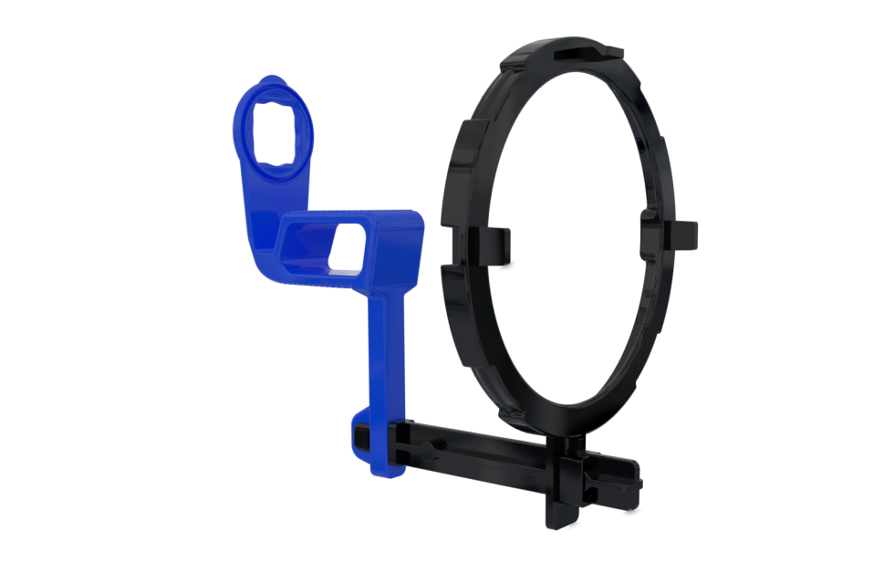
For positioning anterior images.
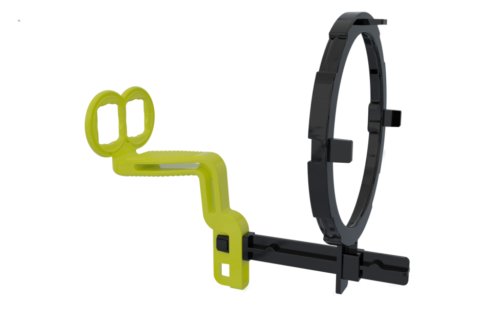
For molar and canine posterior images.
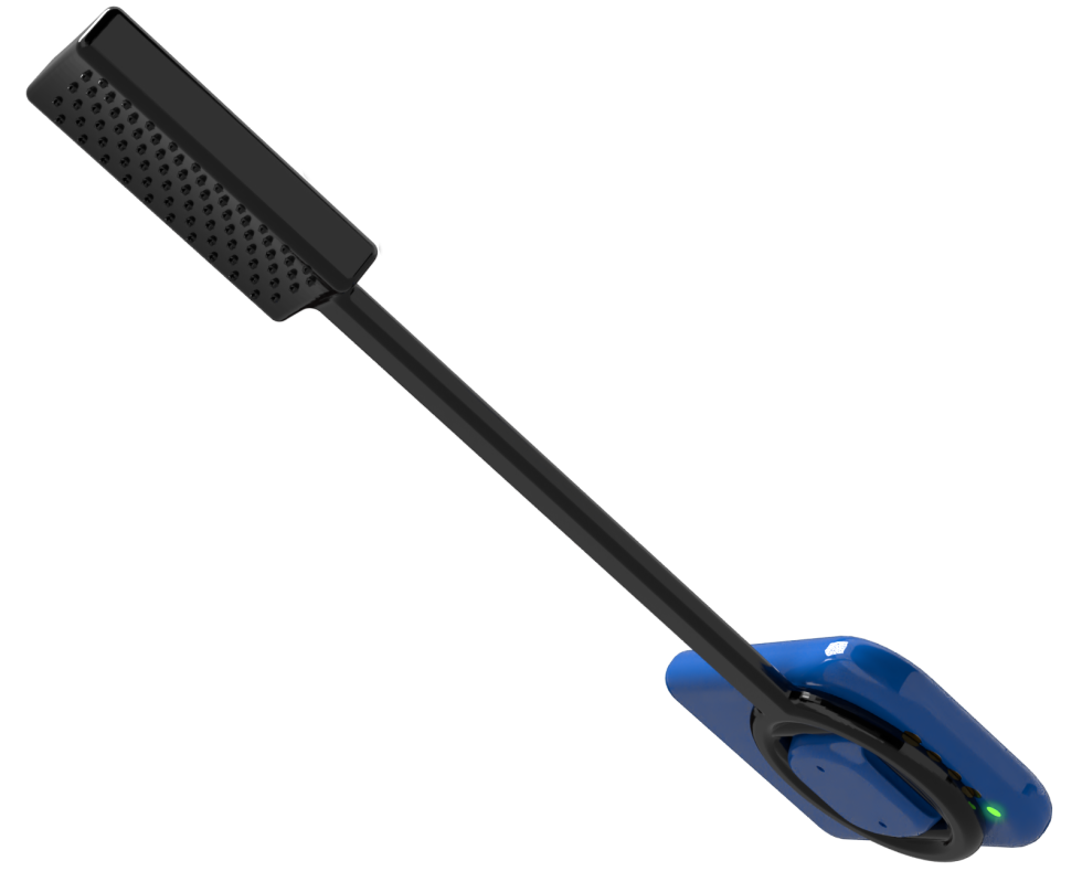
For root canal and endodontic images.
Zero Profile® Comfort System
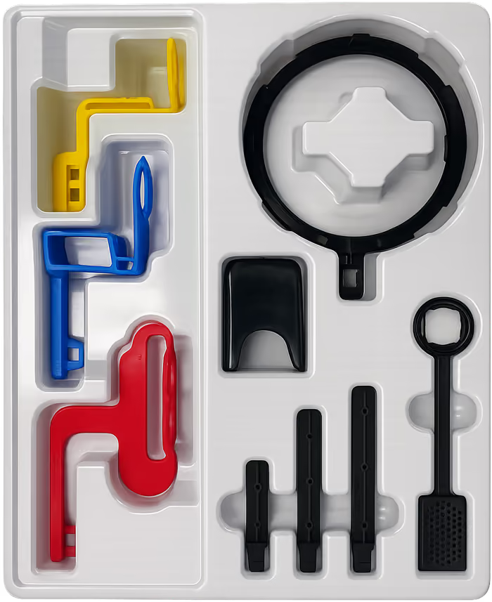
The wireless sensor and low-profile holders minimize intrusion and positioning restrictions so patients can remain more comfortable during imaging.
IP67 water and dust resistance, homogenous silicon construction and a quickly rechargeable battery support daily clinical use.
Soft edges and holder geometry support pediatric patients, sensitive gag reflexes and posterior placements where space is limited.
See it from every side
The source demonstration shows the sensor system, positioning and wireless workflow in use.
DC-Air® integrates with cloud-based and AI-enabled dental environments while preserving the positioning consistency those workflows depend on.
At a glance
The core details behind the wireless imaging system.
Workflow
FTG can help confirm compatibility for your software version and practice configuration.









Head-to-head
Four product decisions distinguish the DC-Air approach from conventional wired and indirect-conversion sensors.
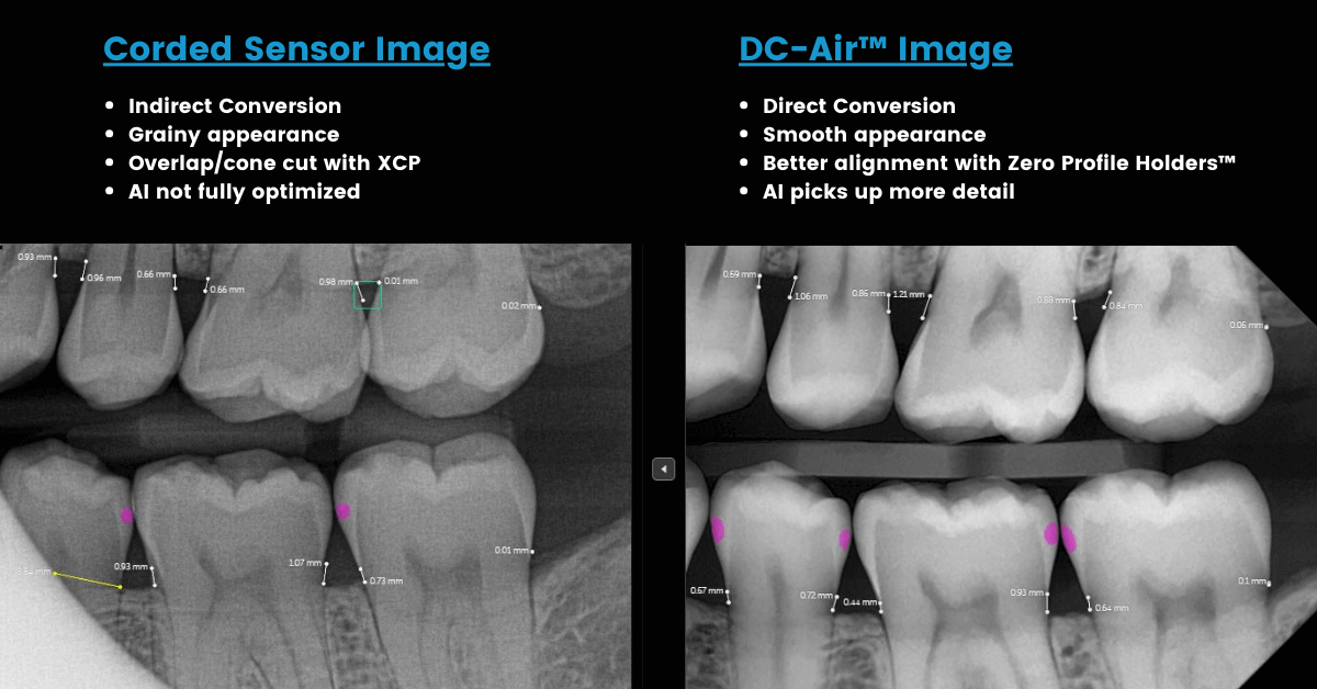
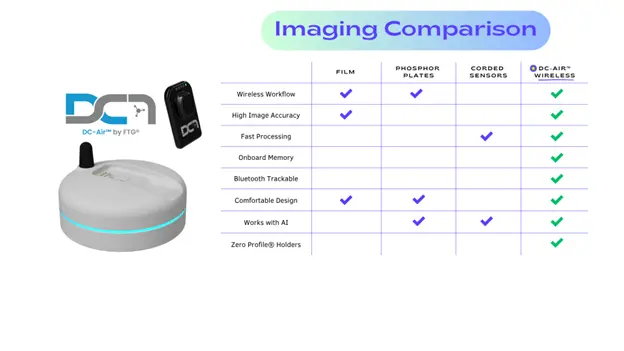
Direct conversion avoids scintillator blur and preserves diagnostic information.
A thinner cable-free profile reduces positioning restrictions.
True wireless operation removes cable management between operatories.
Removing the cable eliminates a common sensor failure point.
Image Accuracy
IA describes how precisely a sensor reproduces the true anatomical structures of teeth, bone and soft tissue with minimal distortion, noise or loss of diagnostic detail.
Visibility of fine details including root tips, caries and trabecular bone.
Differentiation of subtle changes in tissue density.
Minimal distortion or magnification for true-to-scale anatomy.
Less electronic or scatter grain obscuring diagnostic features.
Reliable reproduction across patients and settings.
Accurate image information supports diagnosis, reduces false positives and negatives, and provides a stronger foundation for human and AI interpretation.
Direct-conversion sensors such as DC-Air® convert X-rays directly into an electronic signal without the scintillator and optical-fiber stages that can scatter information. FTG positions that simpler signal path, supported by the MTF comparison above, as the foundation for more accurate diagnostic images.
See it for yourself