
Radiographs
Image Gallery
Clear, concise imaging. All without the hassle of a wire.
Read the evidence yourself
Inspect real DC-Air® captures.
Zoom, pan, invert and adjust brightness, contrast or display sharpening to inspect the available radiographs directly.
Every dentist has a preferred look. Adjust the brightness, contrast and sharpness to match your viewing preference, while preserving the IA (Image Accuracy) of the DC-Air® True Wireless® sensor.
Use plus and minus to zoom, arrow keys to pan, bracket keys to switch images, and Home to reset.
Loading image
Drag to pan once zoomed, pinch or use the controls to zoom, and select a thumbnail to change images. Display adjustments are non-destructive and do not modify the source radiograph.
Found your preferred look?
Send us your settings.
These are the same sliders as the DCR TWAIN capture software. Share the look you dialed in and FTG will configure your chairside setup to match it exactly.
This is the real workflow
You’re already using our TWAIN flexibility.
The controls above work just like DCR TWAIN capture software chairside: invert, adjust and inspect without touching the source image. See it live on your own operatory screen.
Same exposure. Two technologies.
Compare direct conversion with a conventional sensor.
Move the divider to examine the source comparison supplied with the DC-Air landing page.
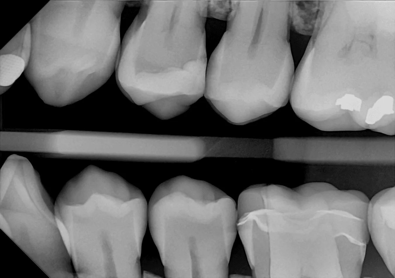
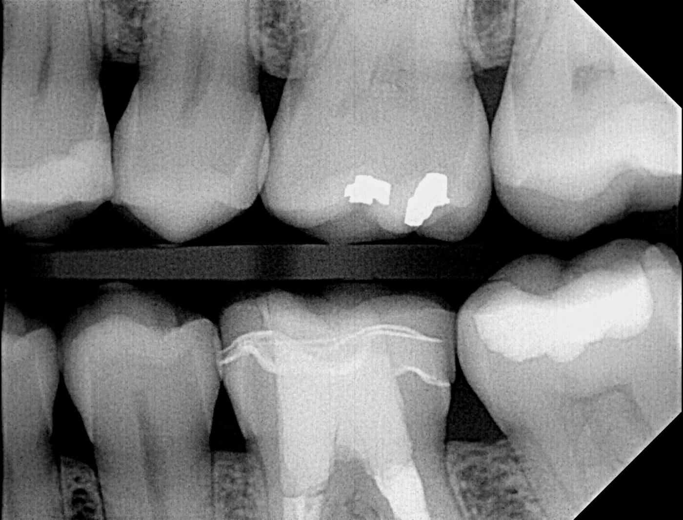
Drag the divider, use the arrow keys, or select a point along the image.
See the clear difference.
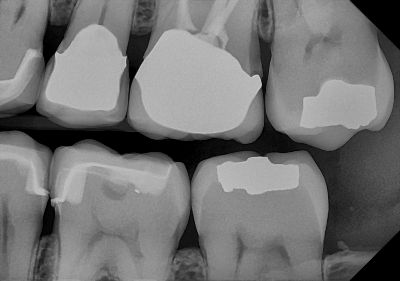
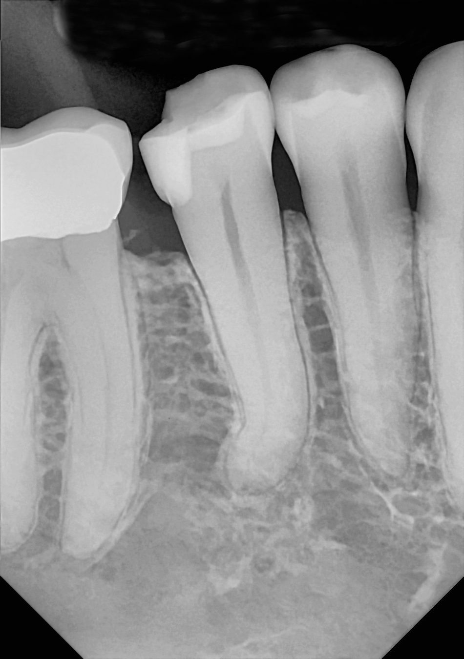
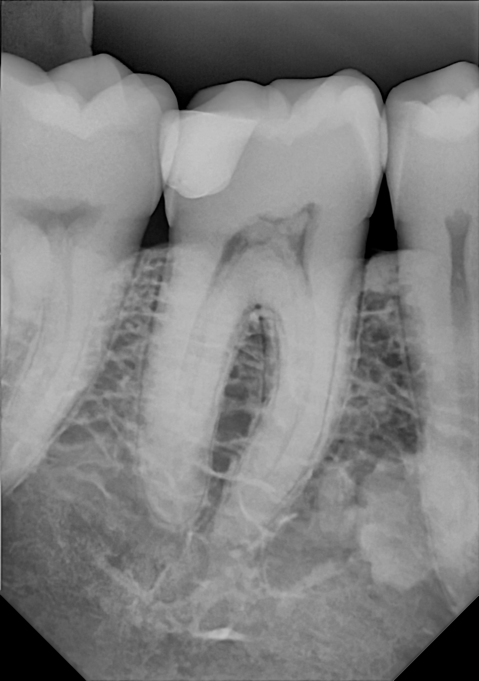
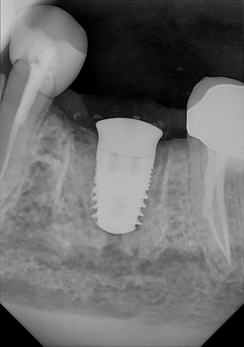
Only DC-Air® features Direct CMOS technology that creates images directly from X-ray photons. True clarity is achieved without relying on sharpening enhancements.
The images in this gallery are from actual DC-Air® users. The only adjustments applied to some images are brightness or contrast.
Compare DC-Air® with your current sensors, PSP plates or film through FTG’s clinical evaluation program.
Discuss an evaluationImage gallery
Bitewings
Comfortably position the rounded edges of the sensor toward the mesial or distal aspects of the mouth depending on the anatomy you want to see.
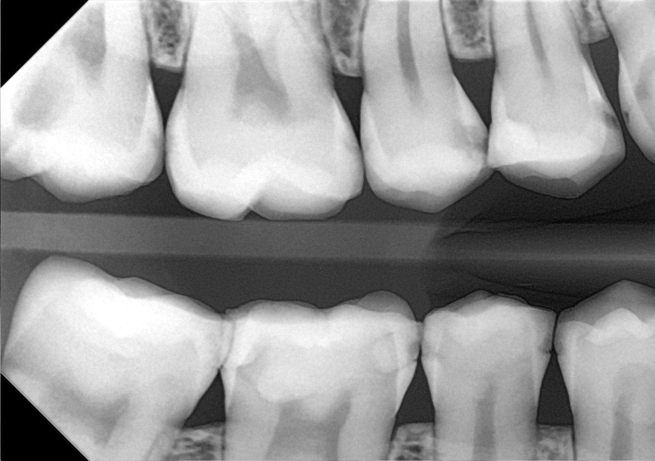
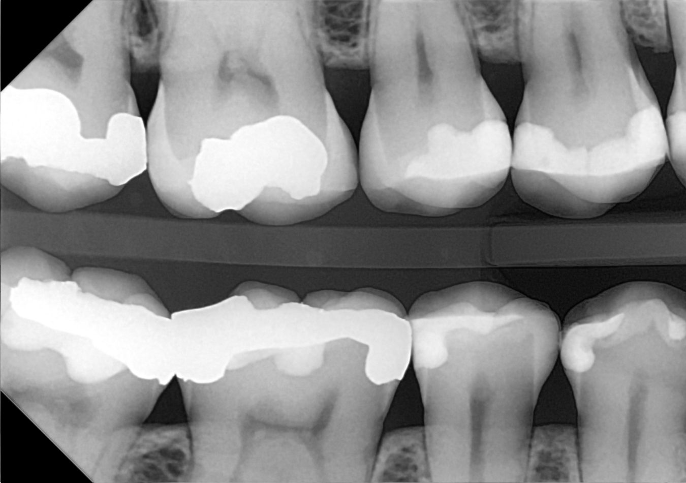
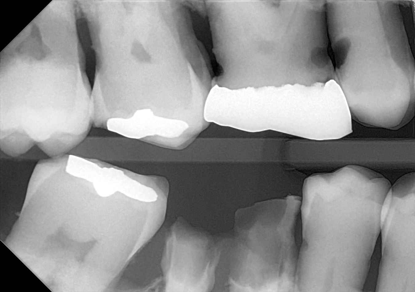
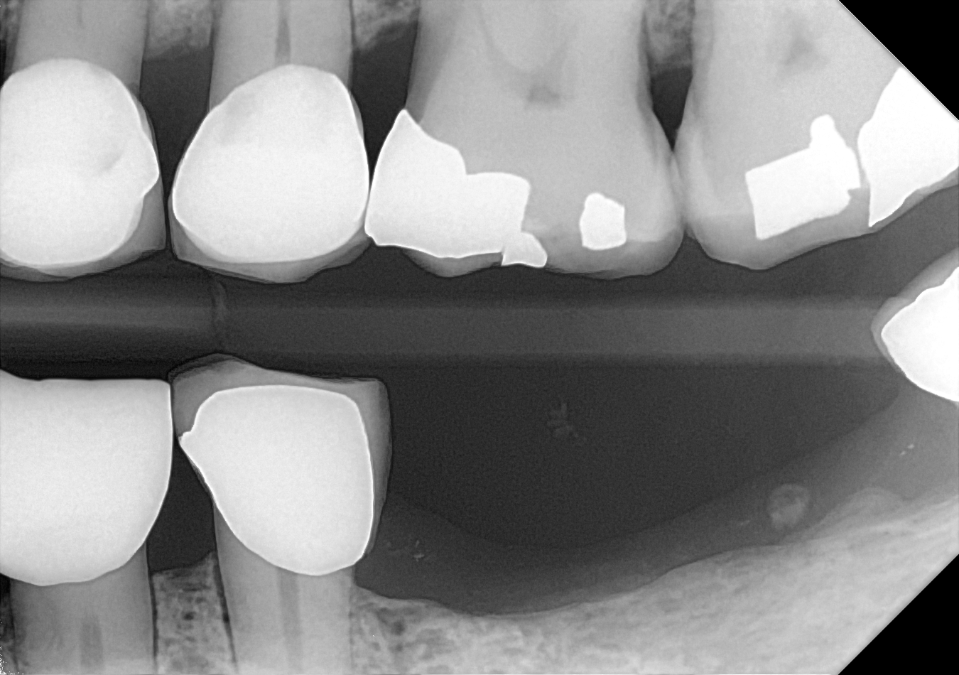
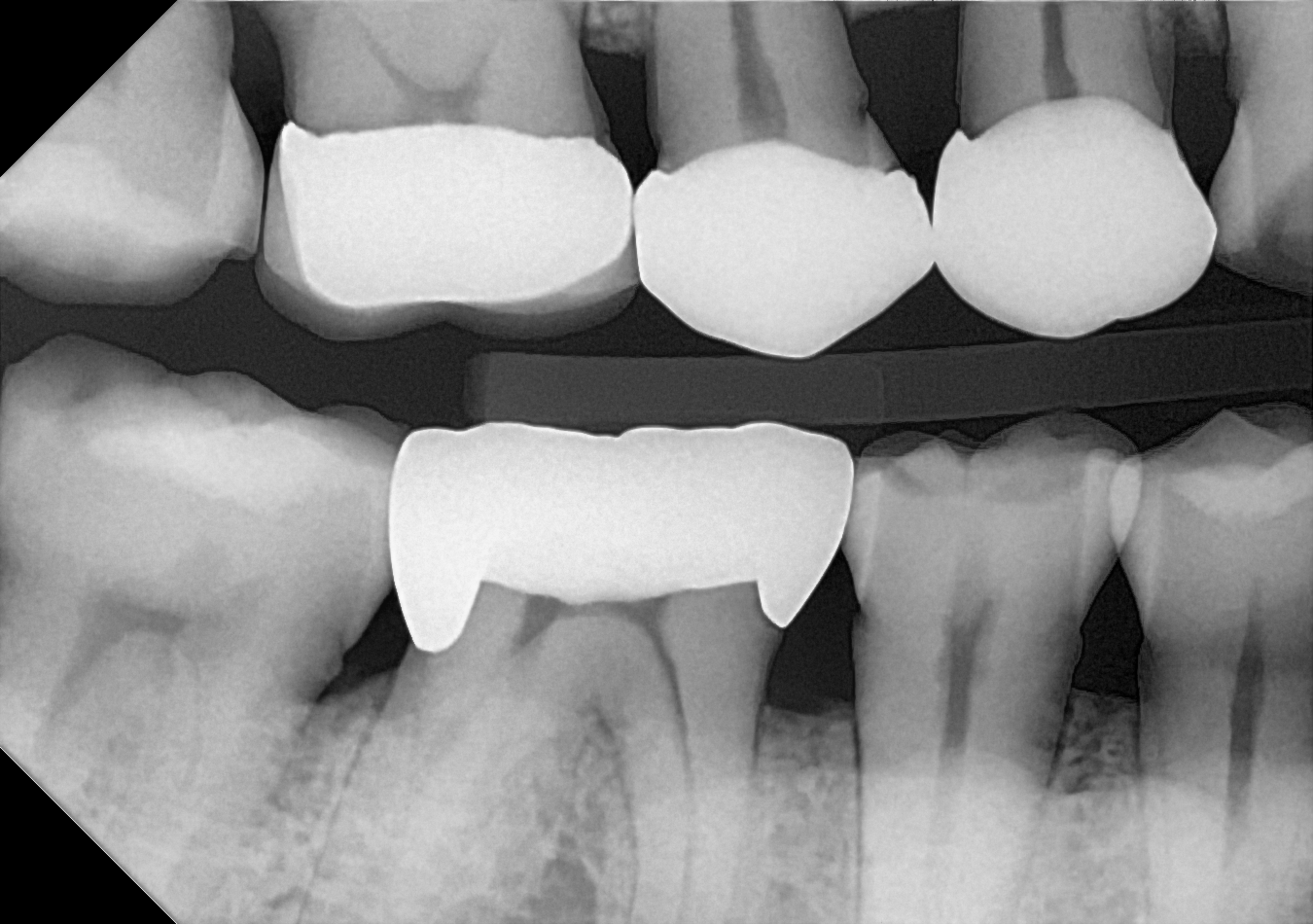
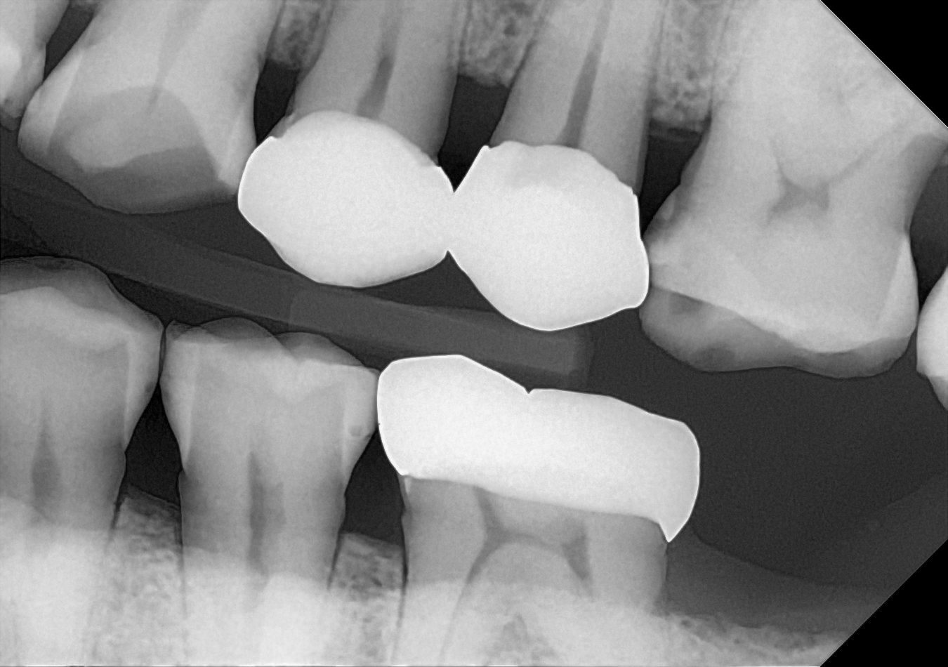
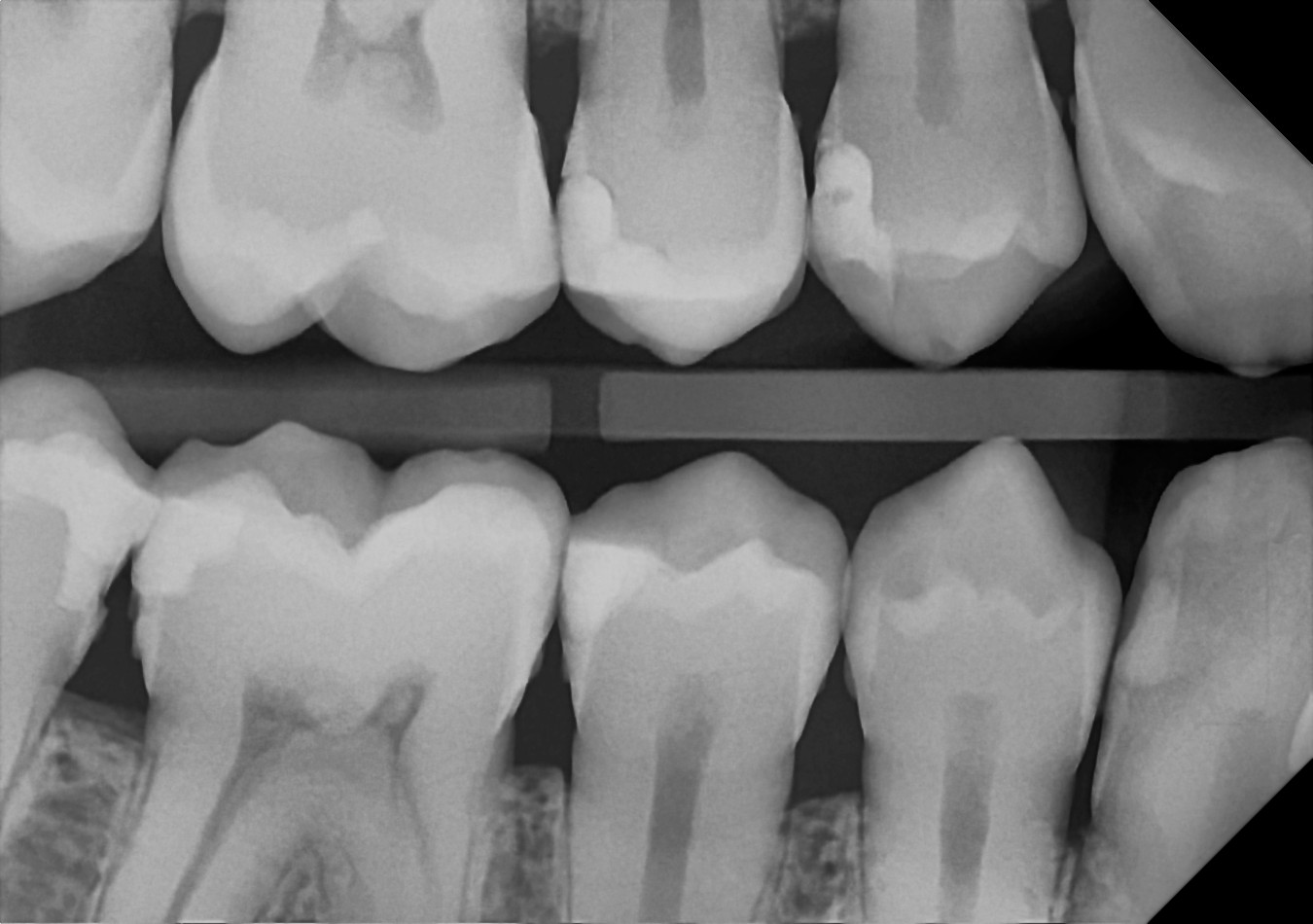
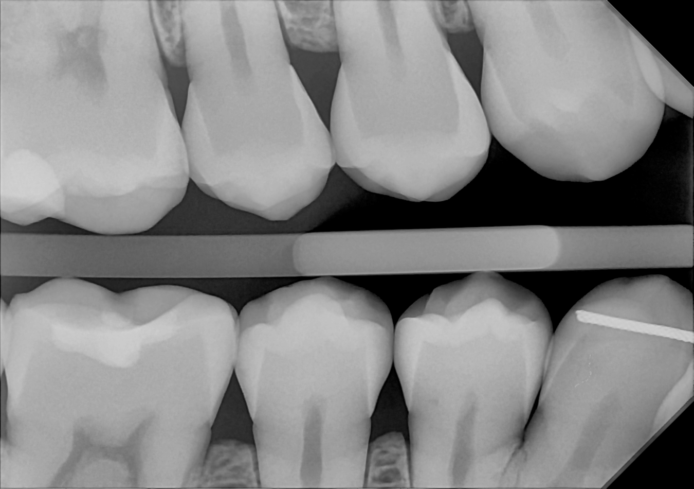
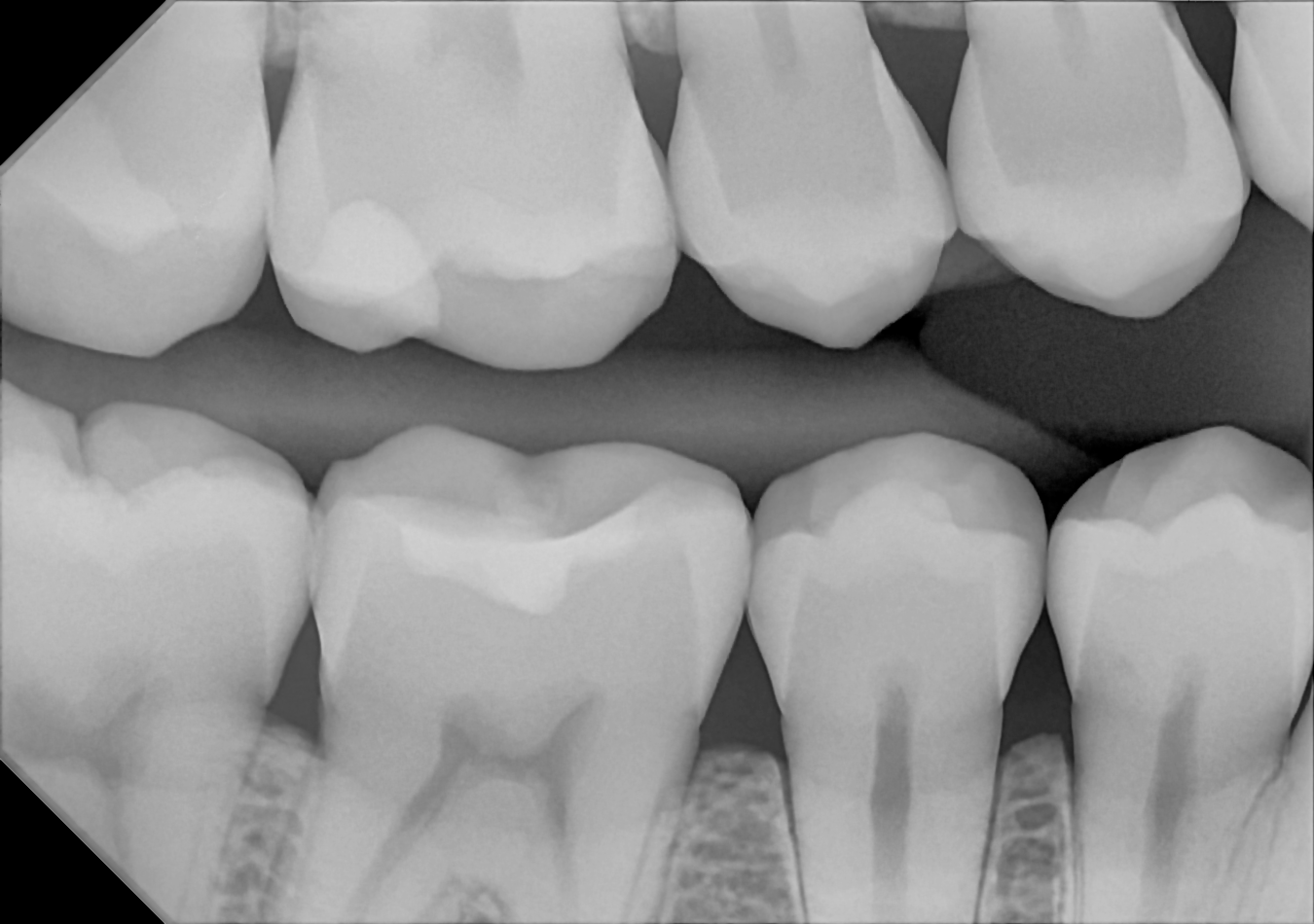
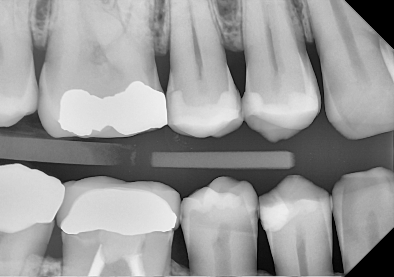
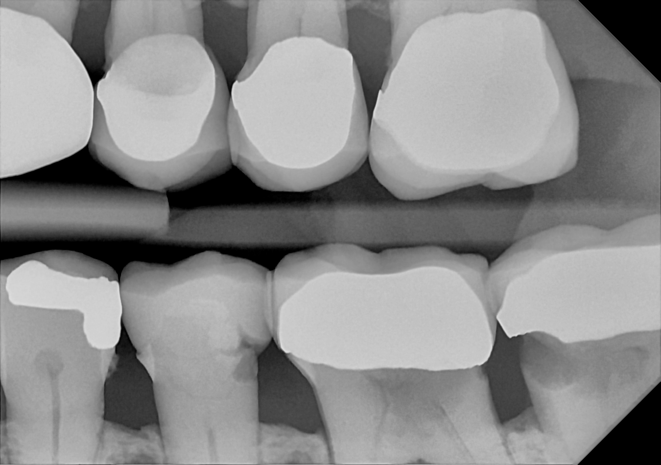
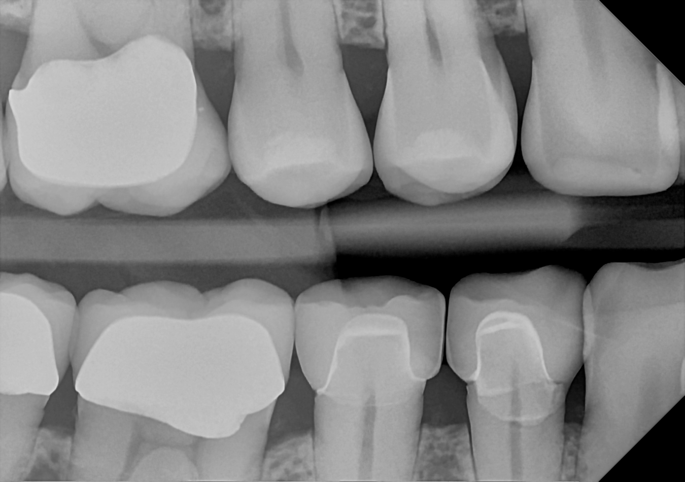
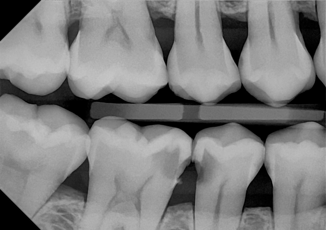
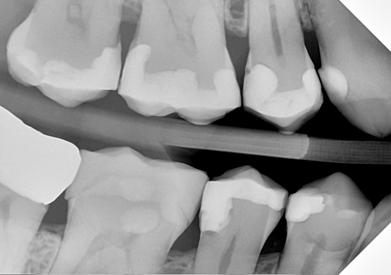
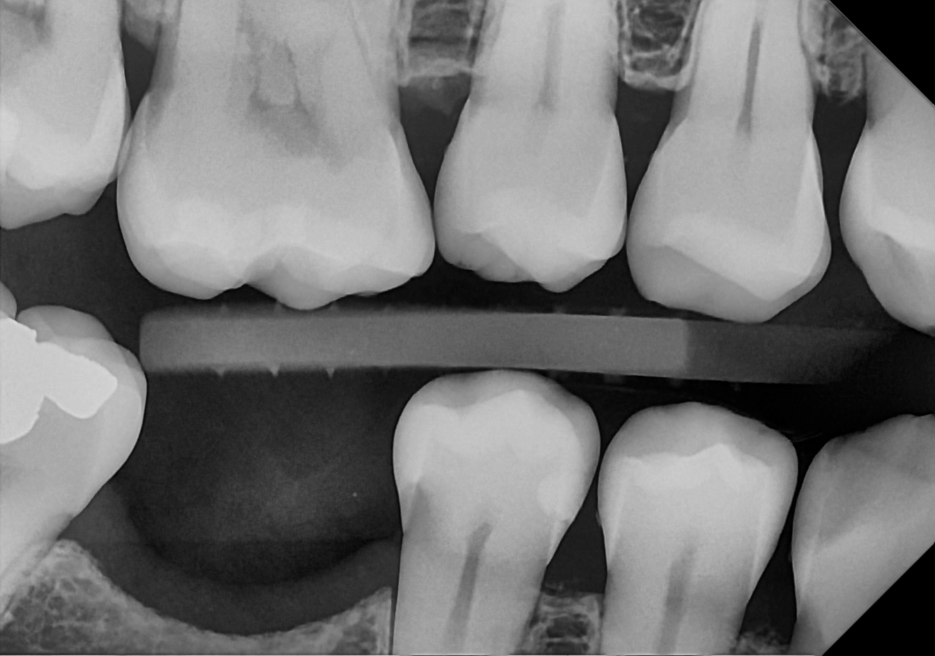
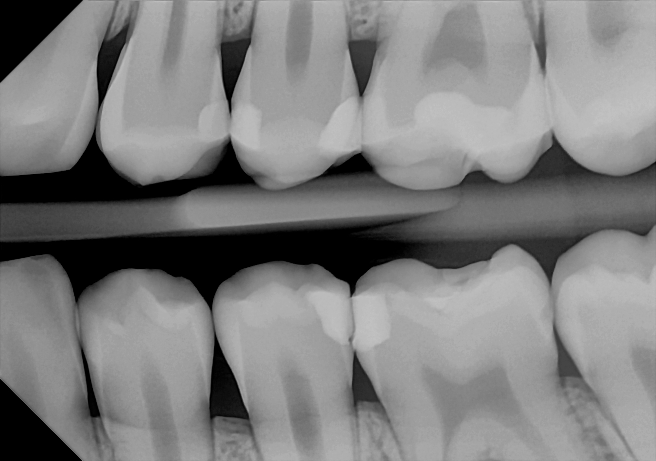
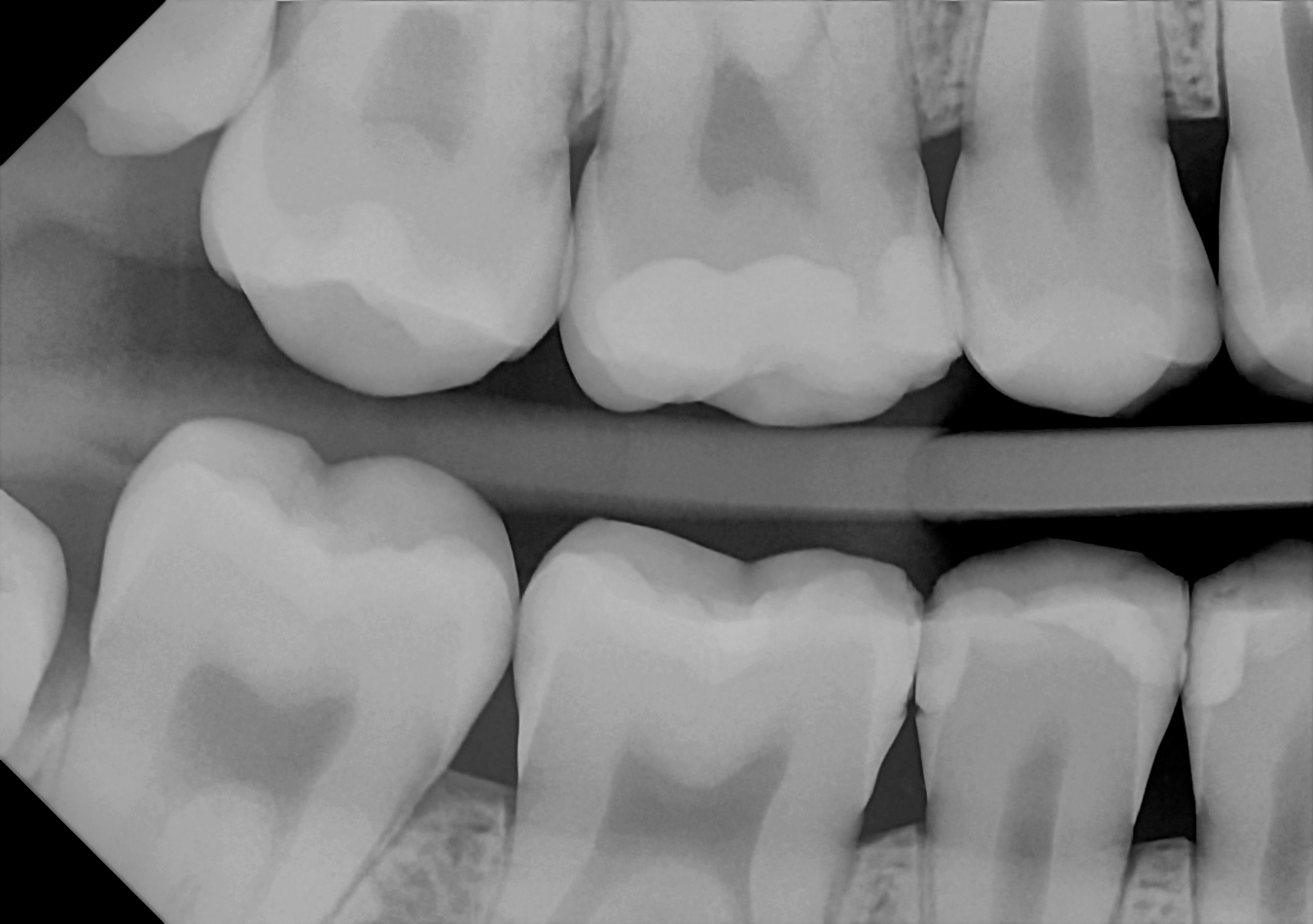
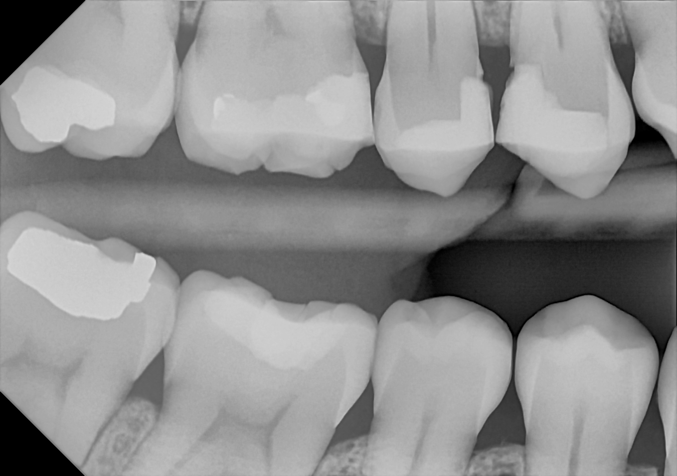
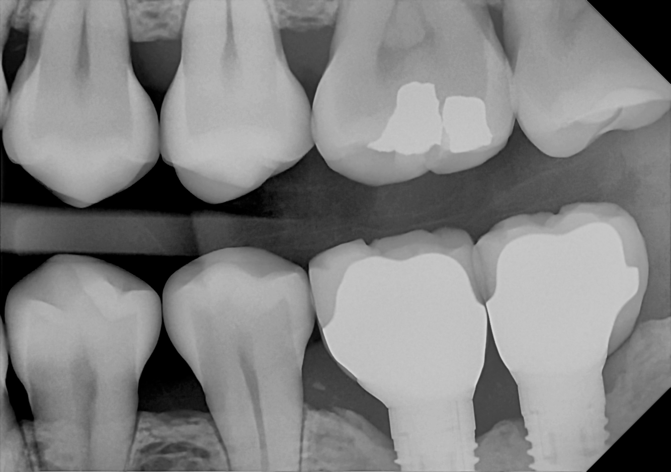
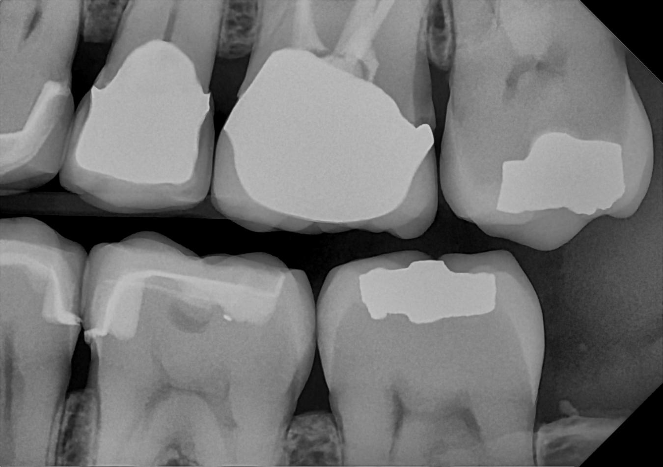
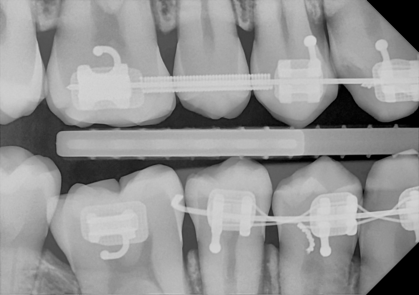
Image gallery
Anterior PAs
Take PAs with ease using the anterior holder, posterior holder, occlusal sheath or specialty endodontic positioning device.
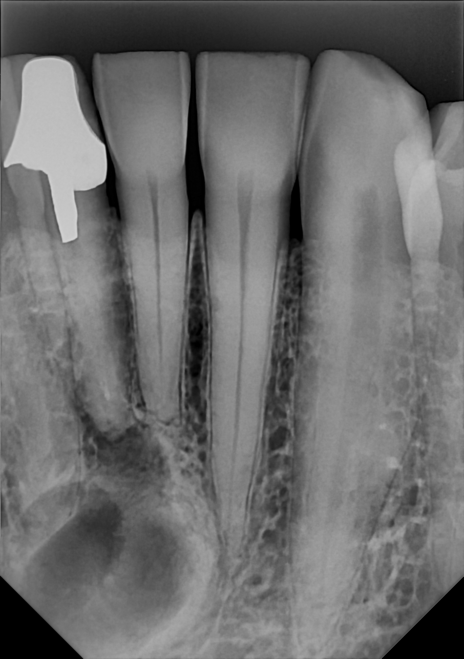
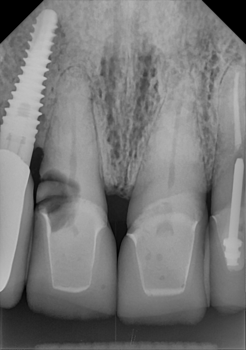
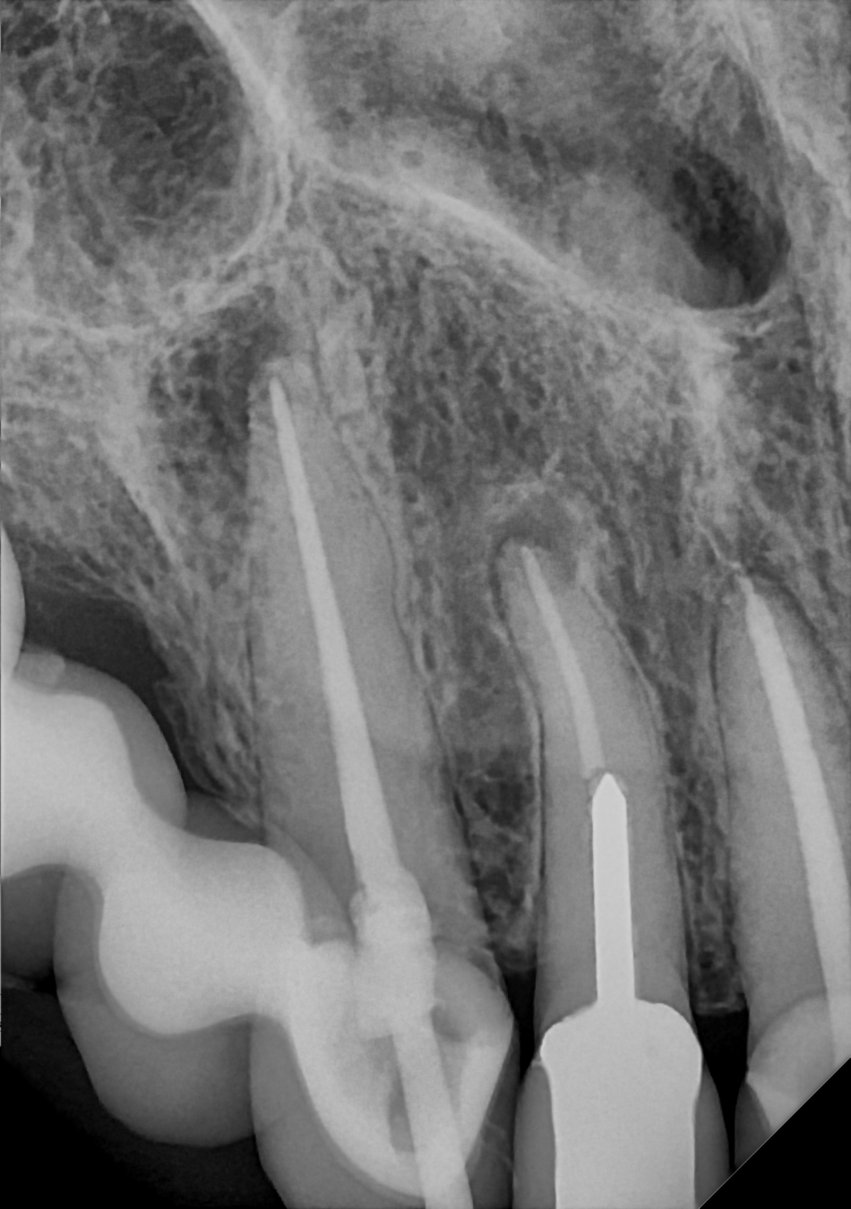
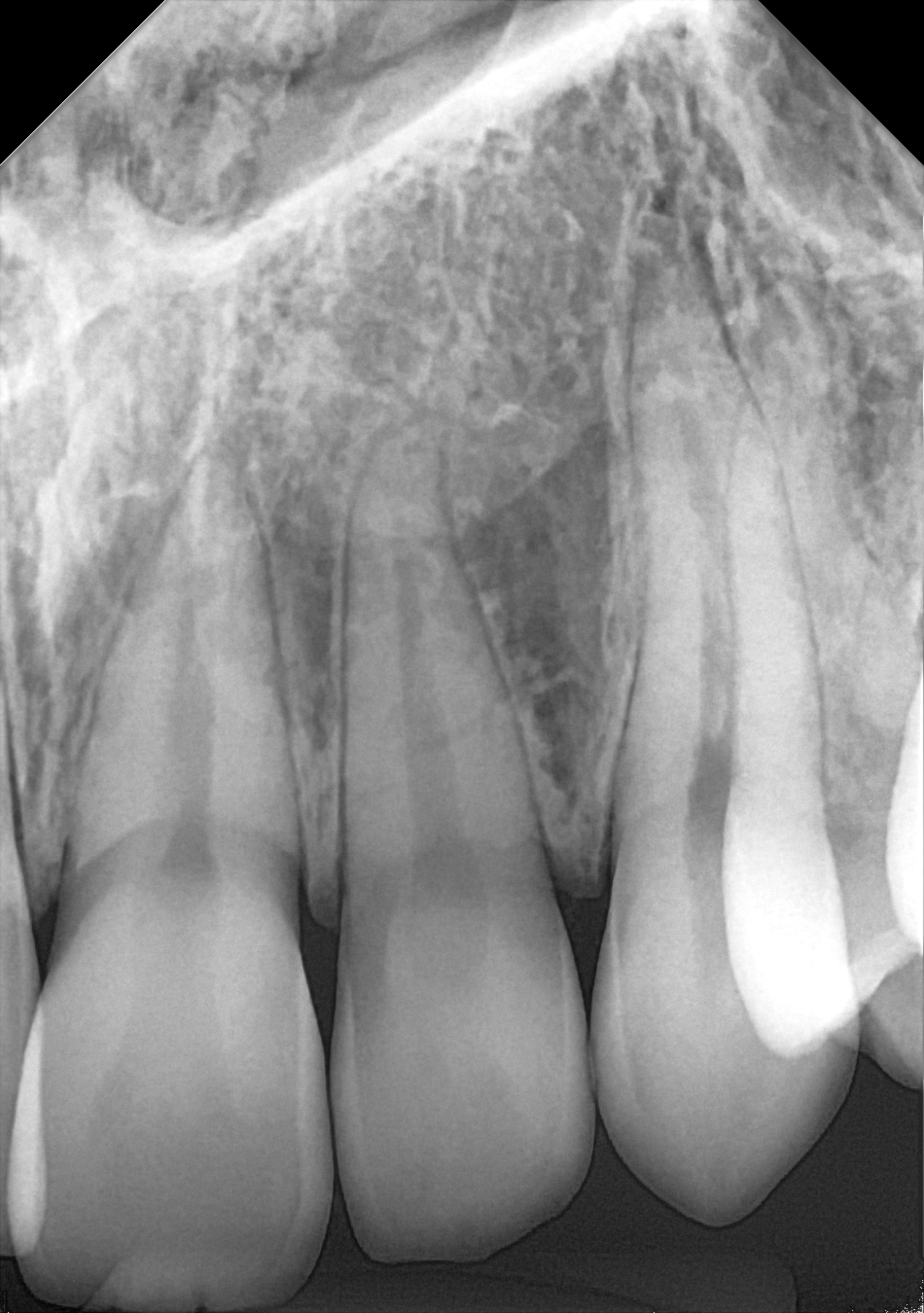
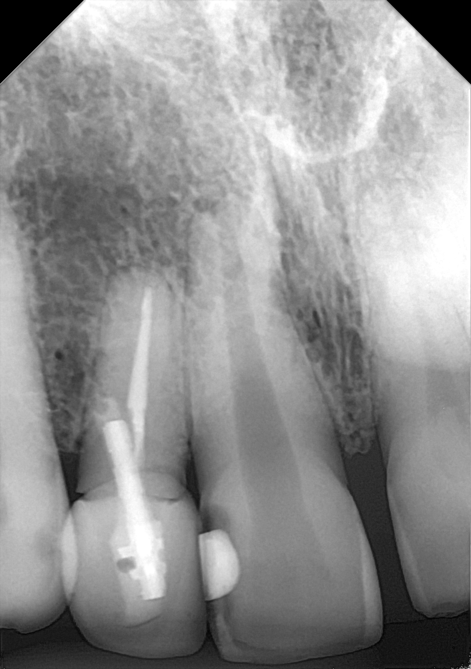
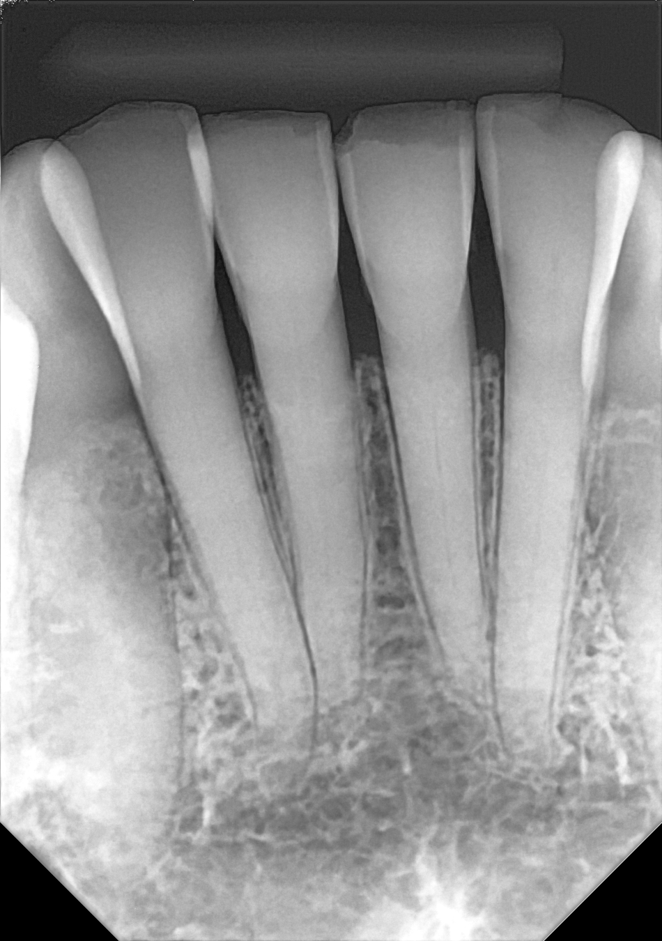
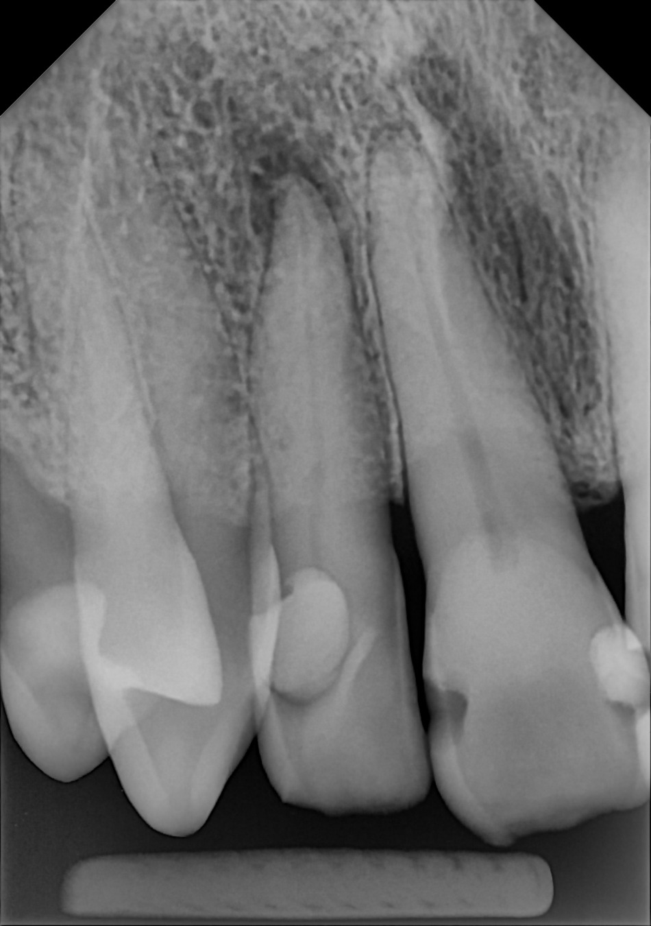
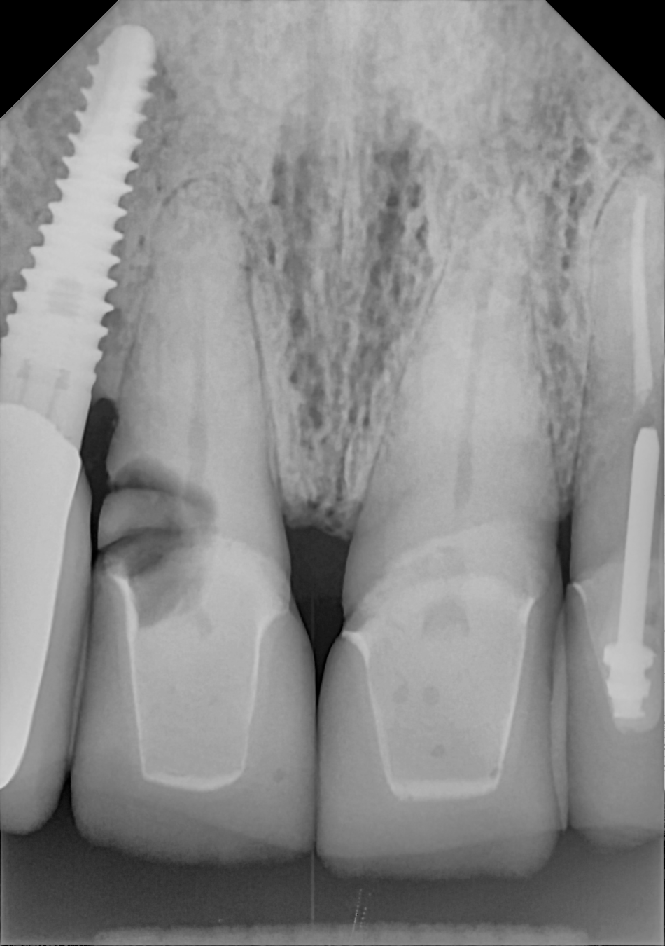
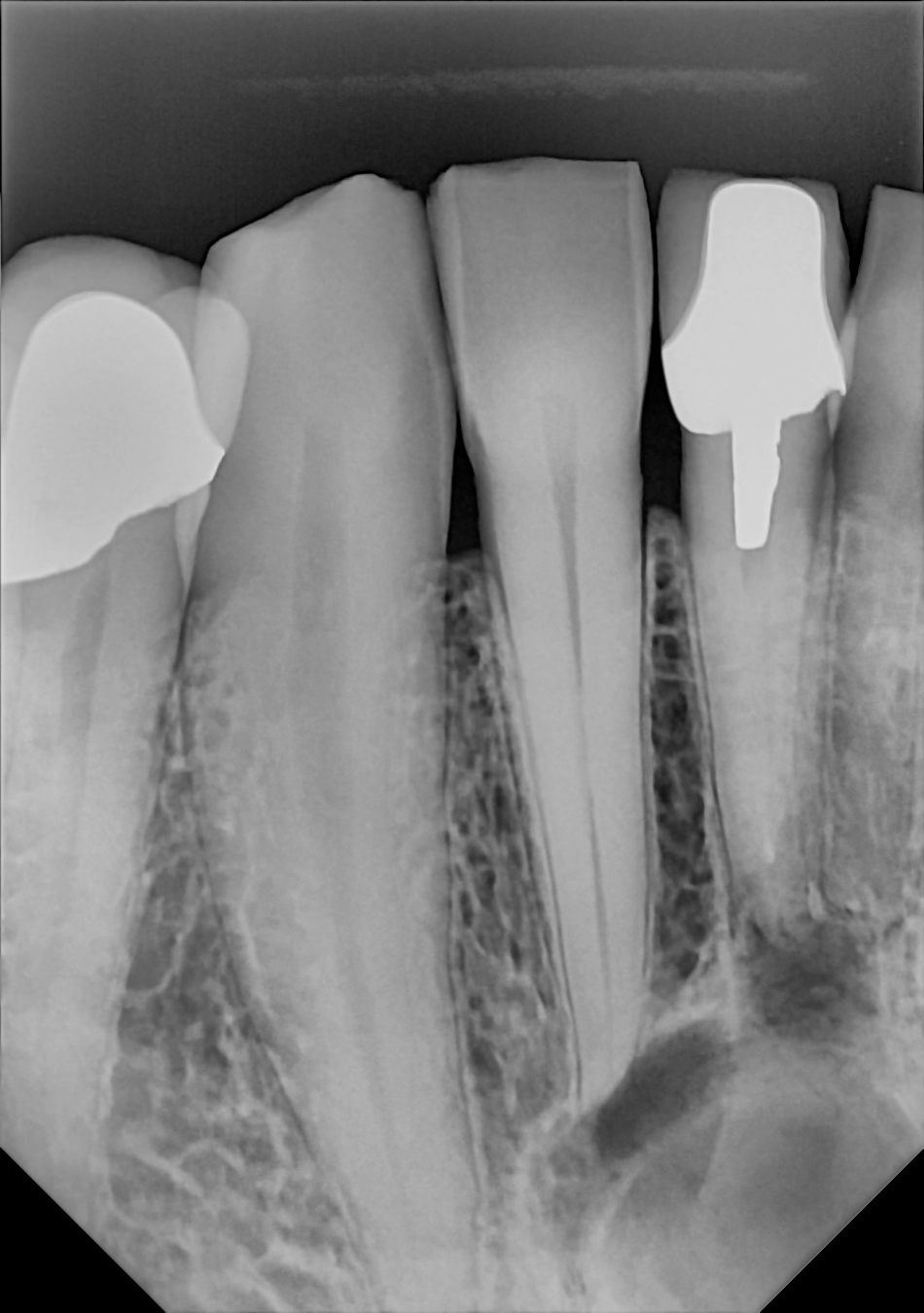
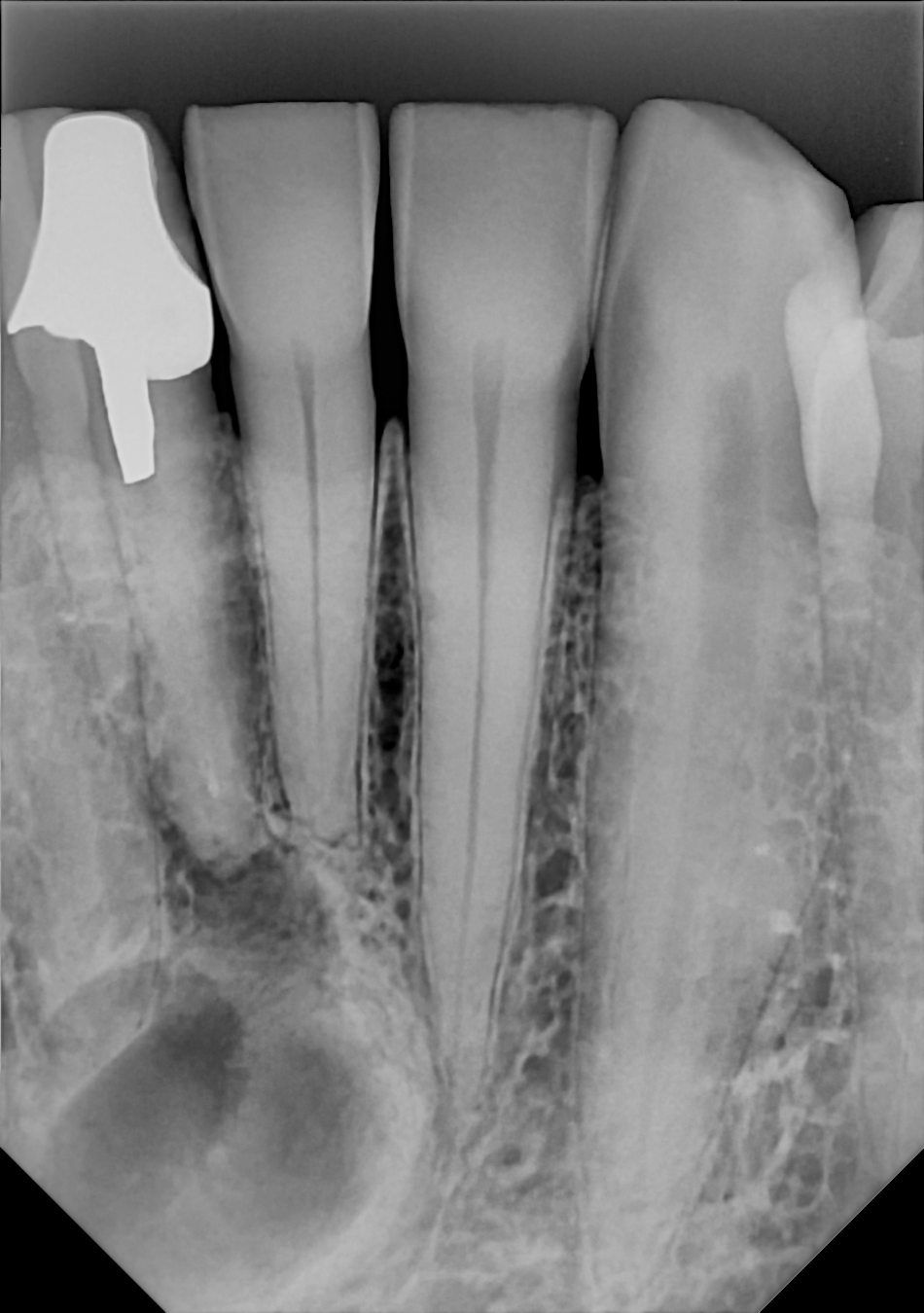
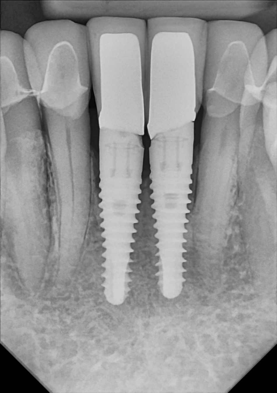
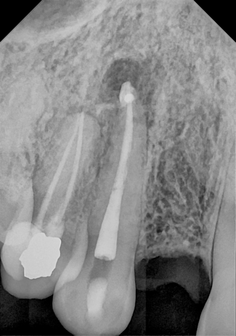
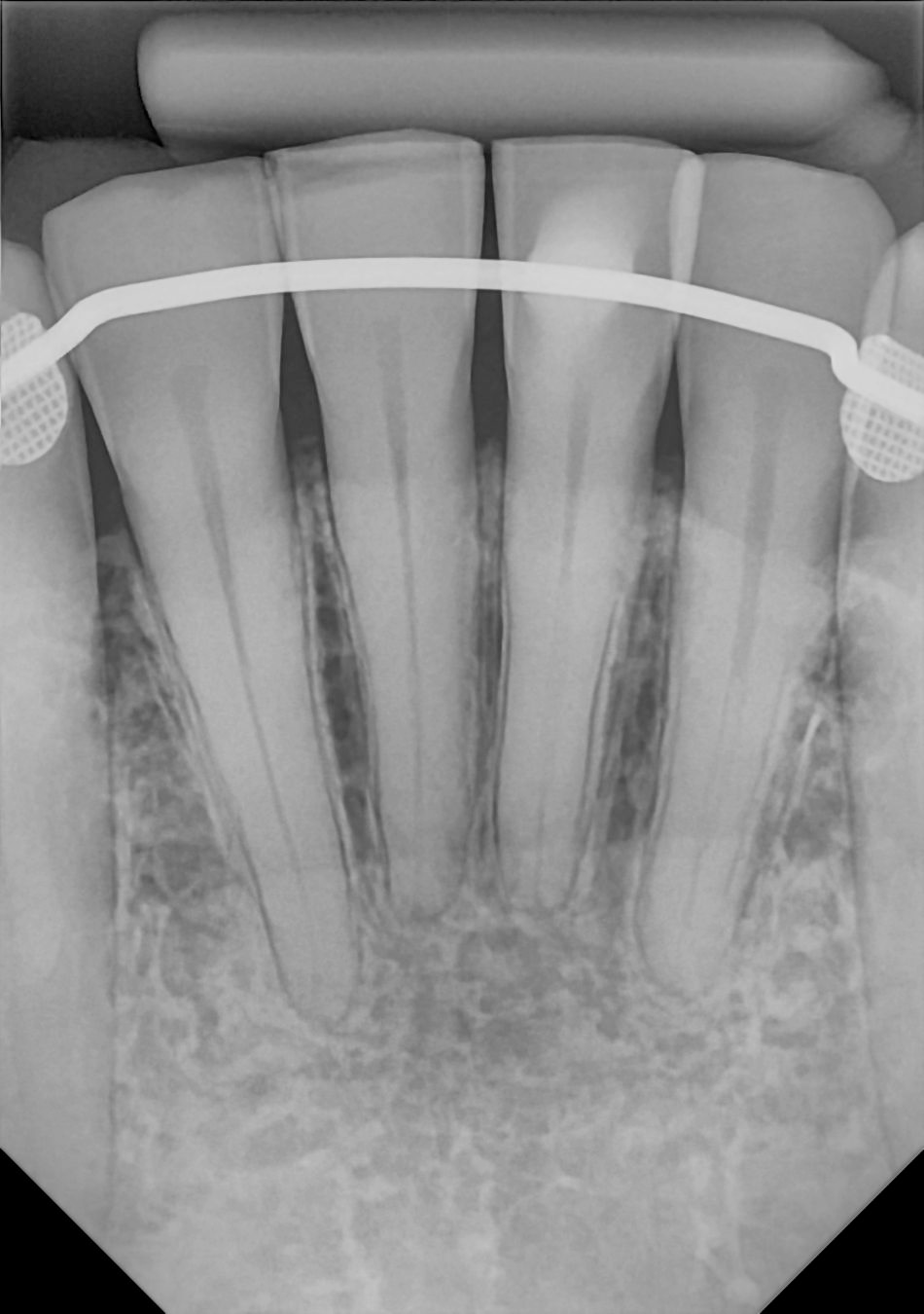
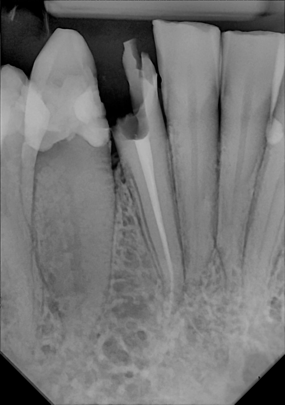
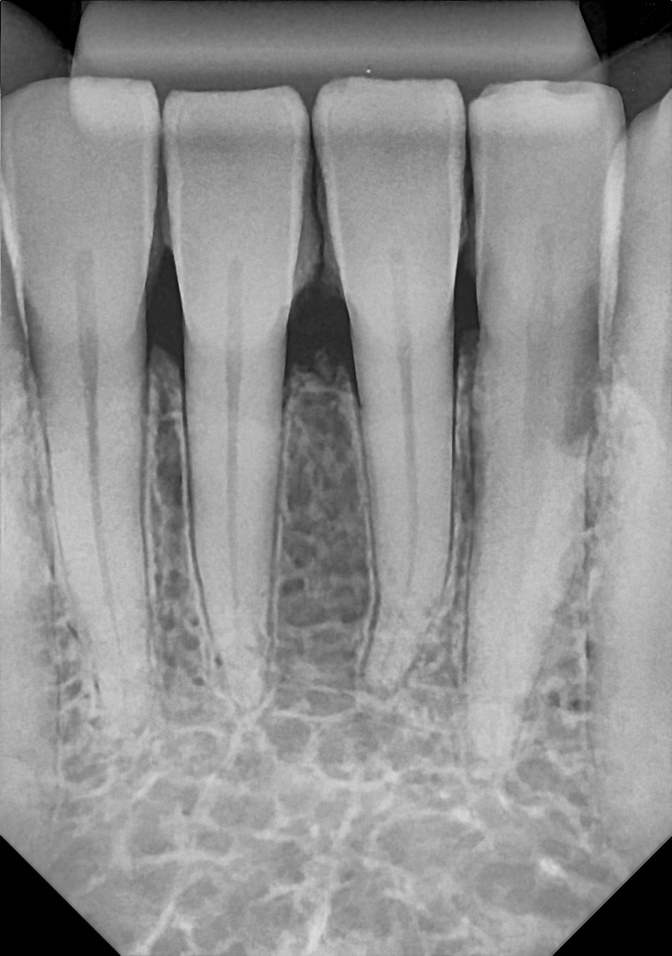
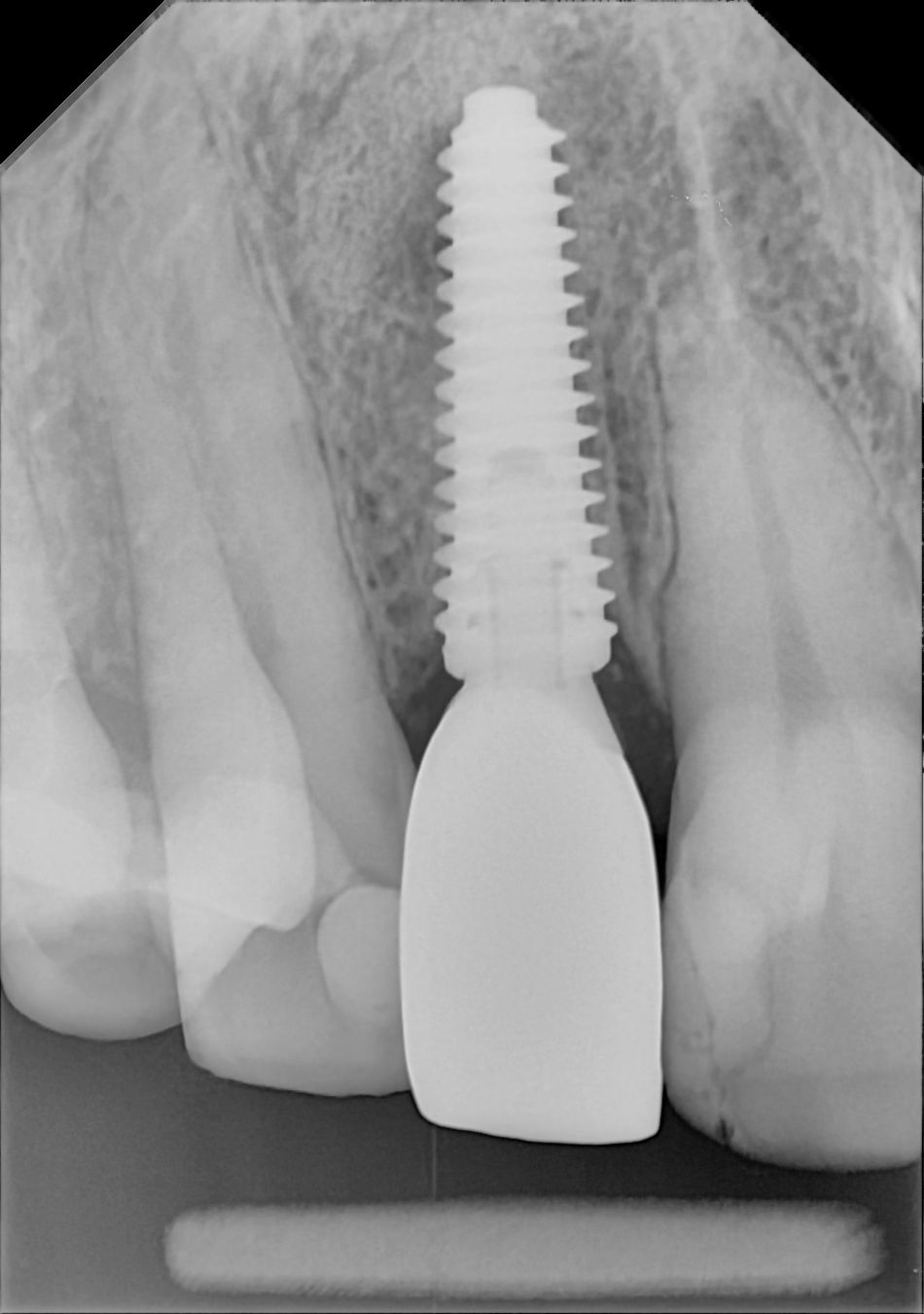
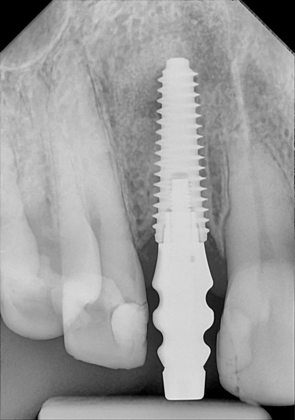
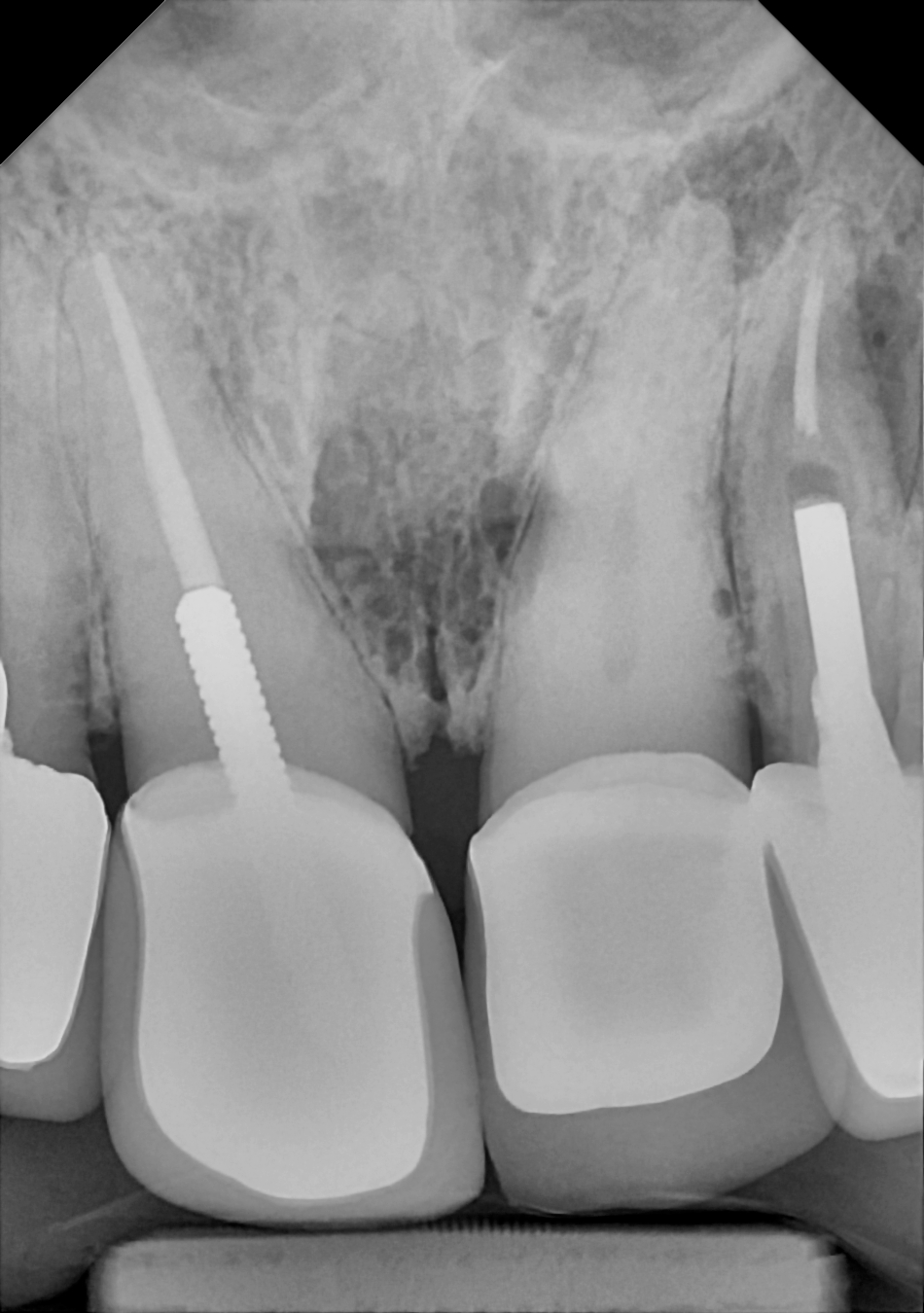
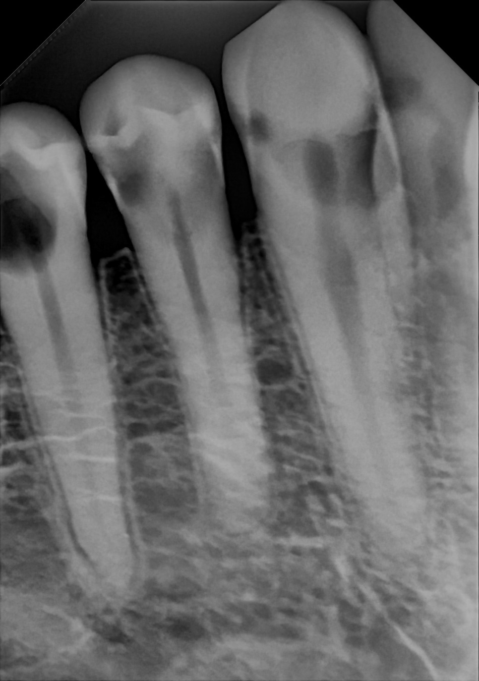
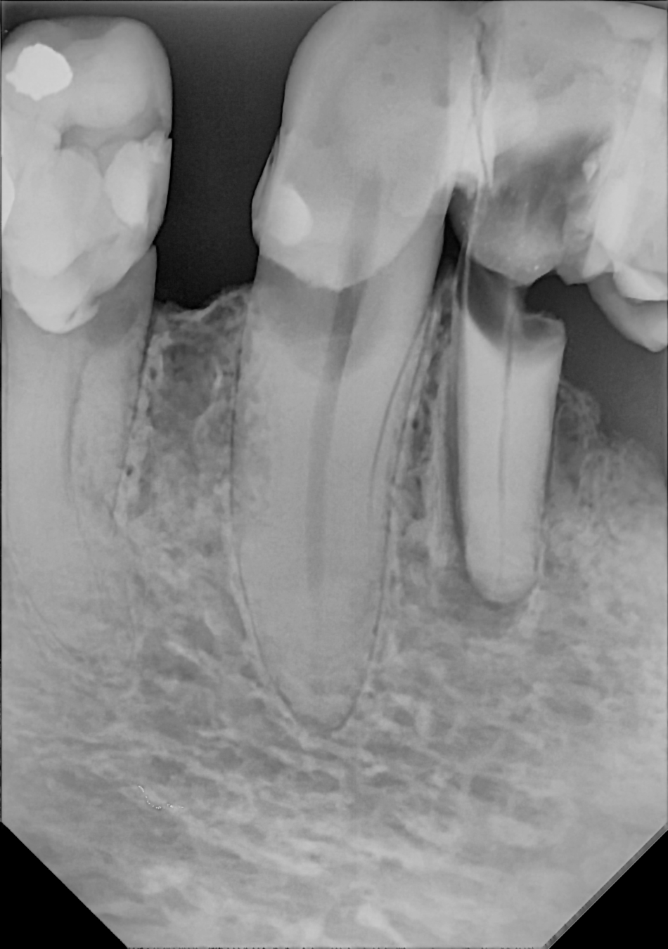
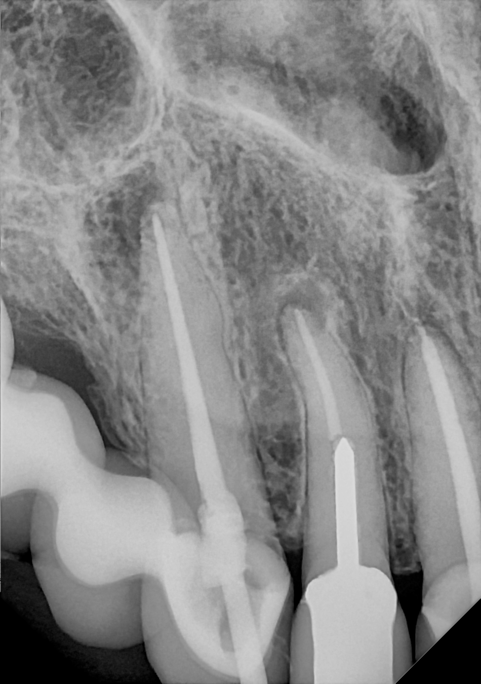
Image gallery
Posterior PAs
Take PAs with ease using the anterior holder, posterior holder, occlusal sheath or specialty endodontic positioning device.
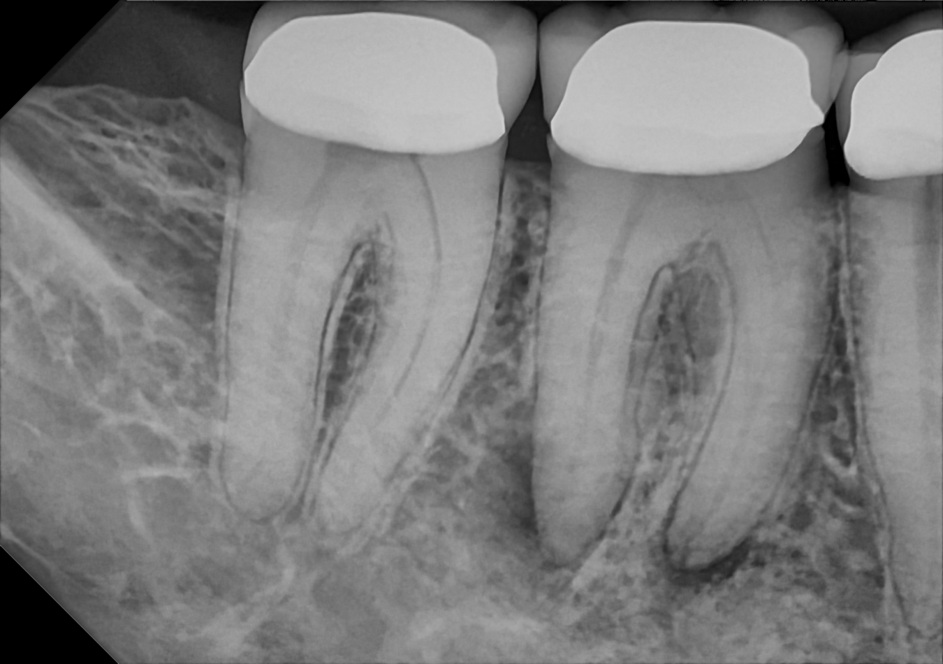
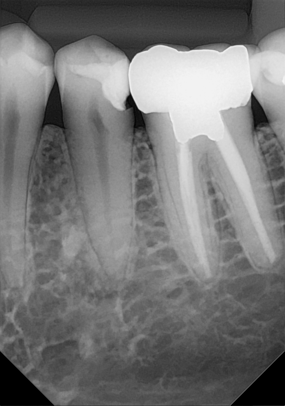
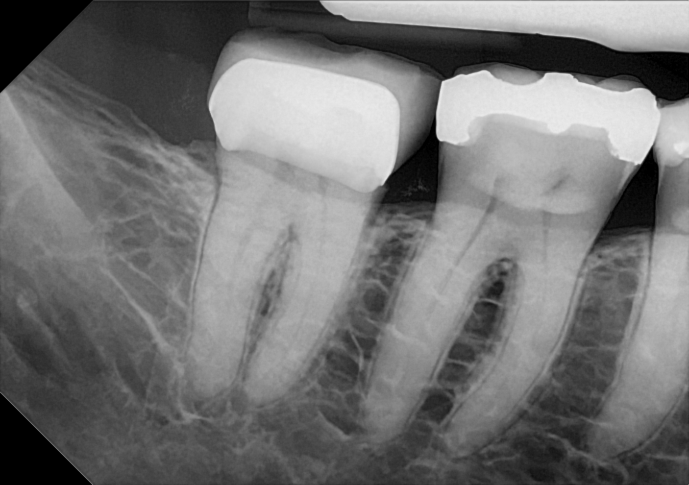
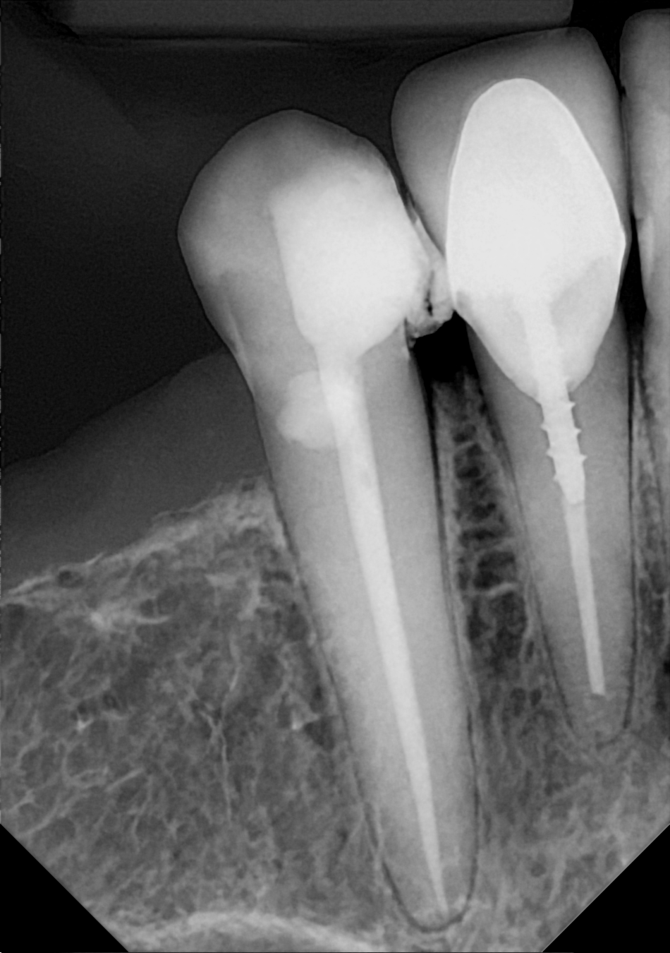
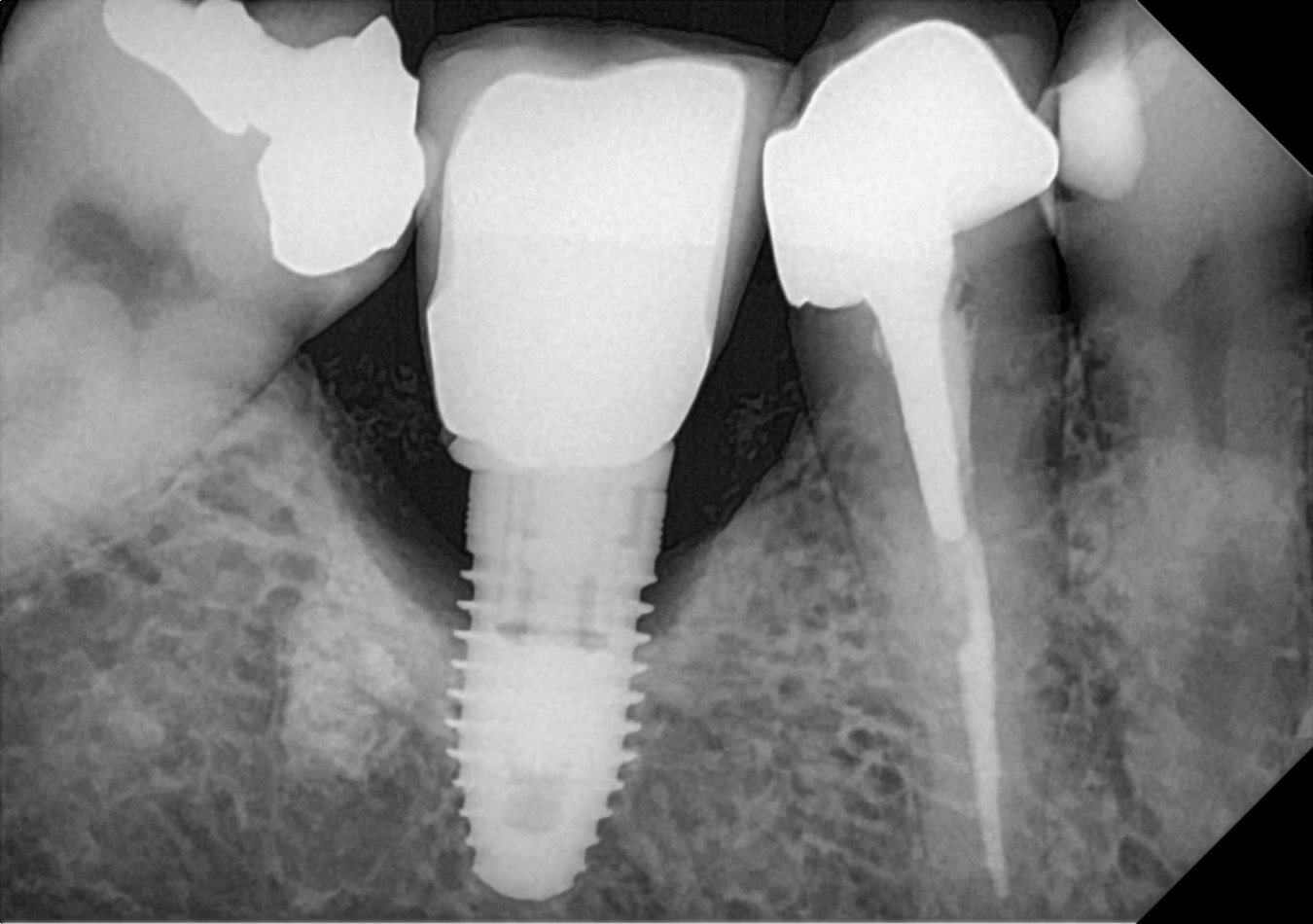
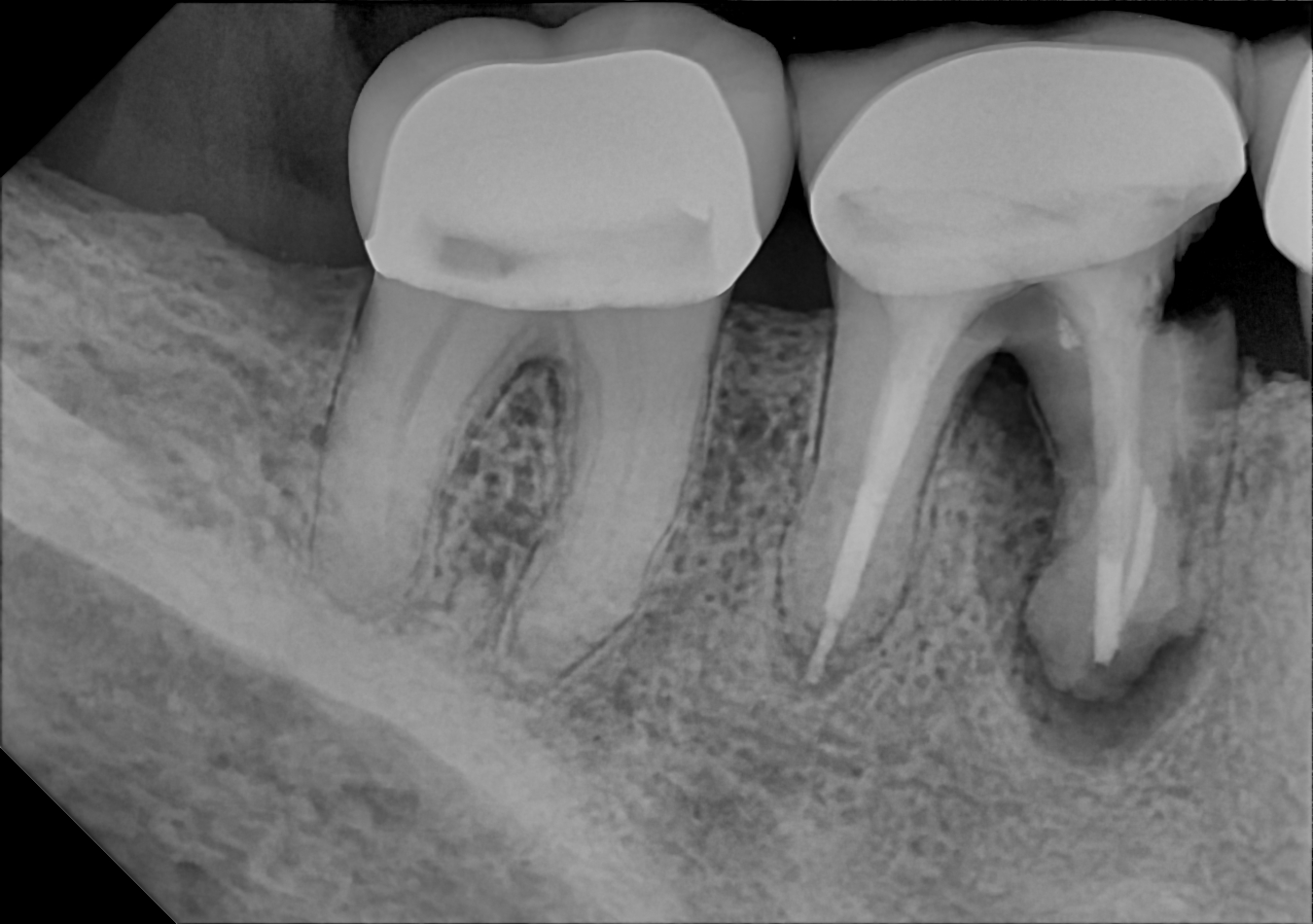
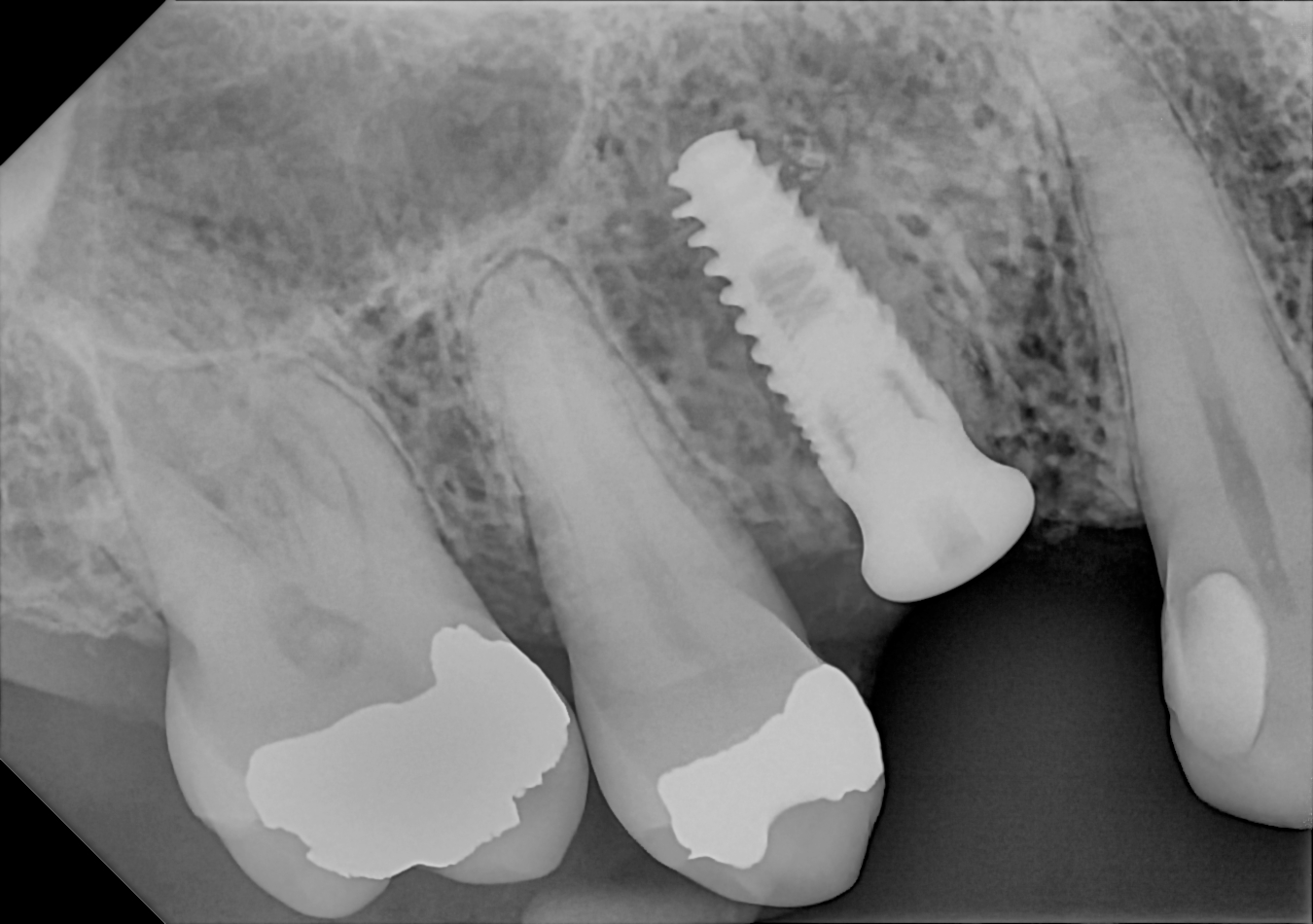
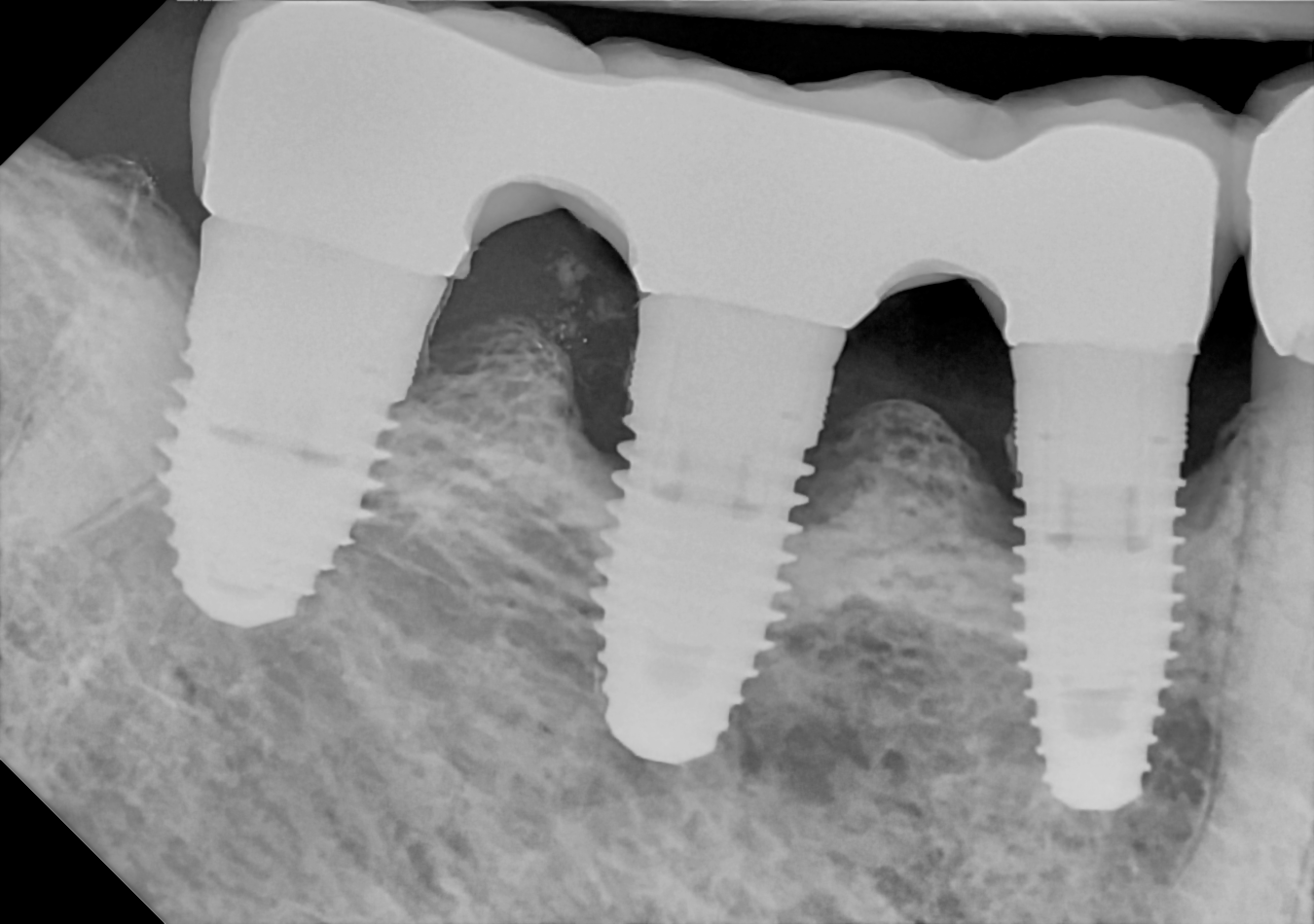
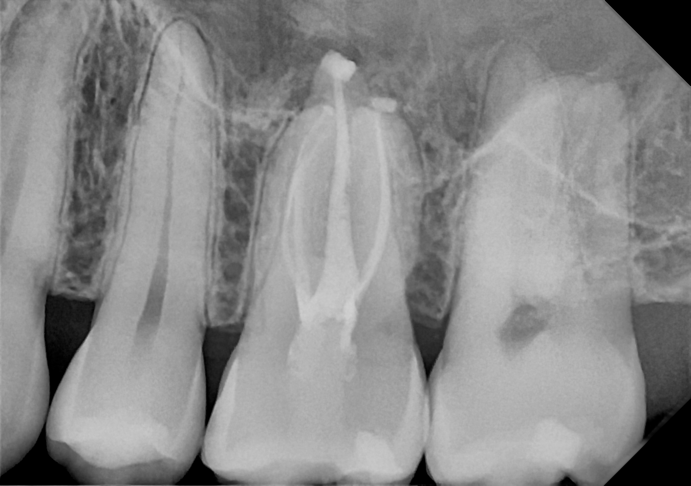
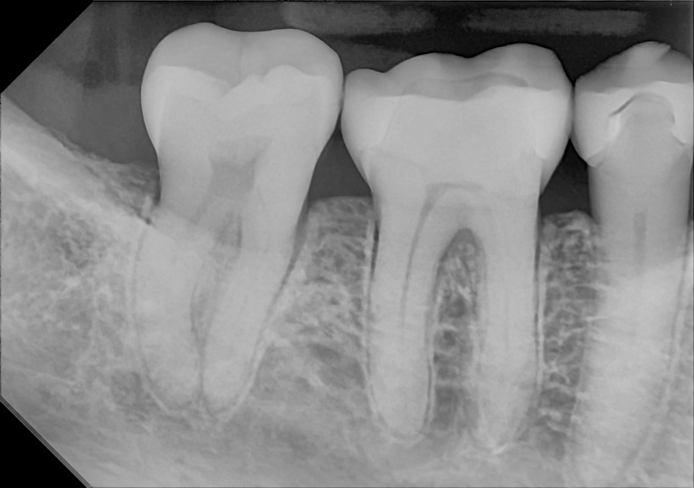
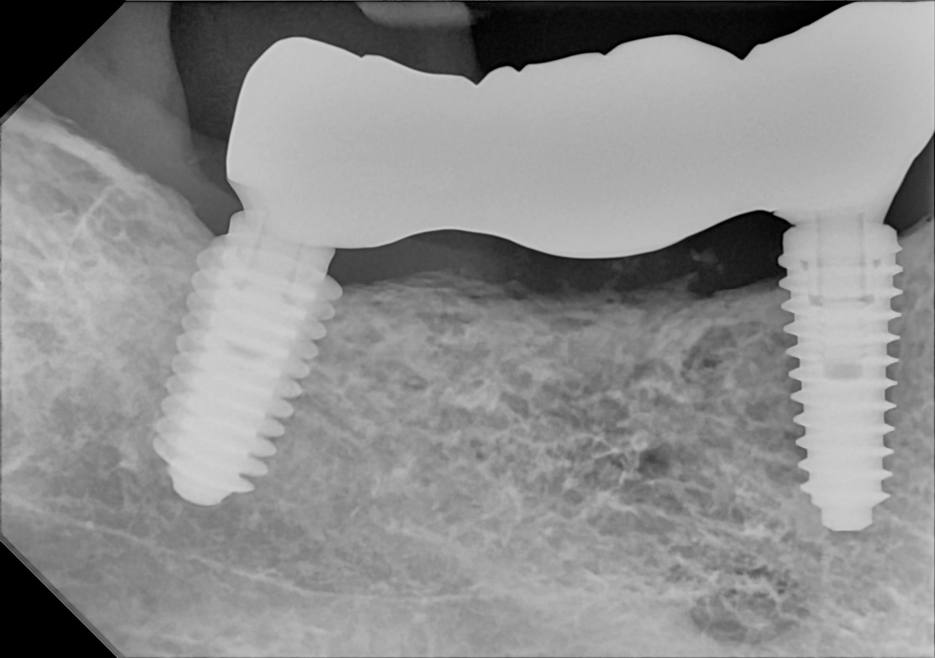
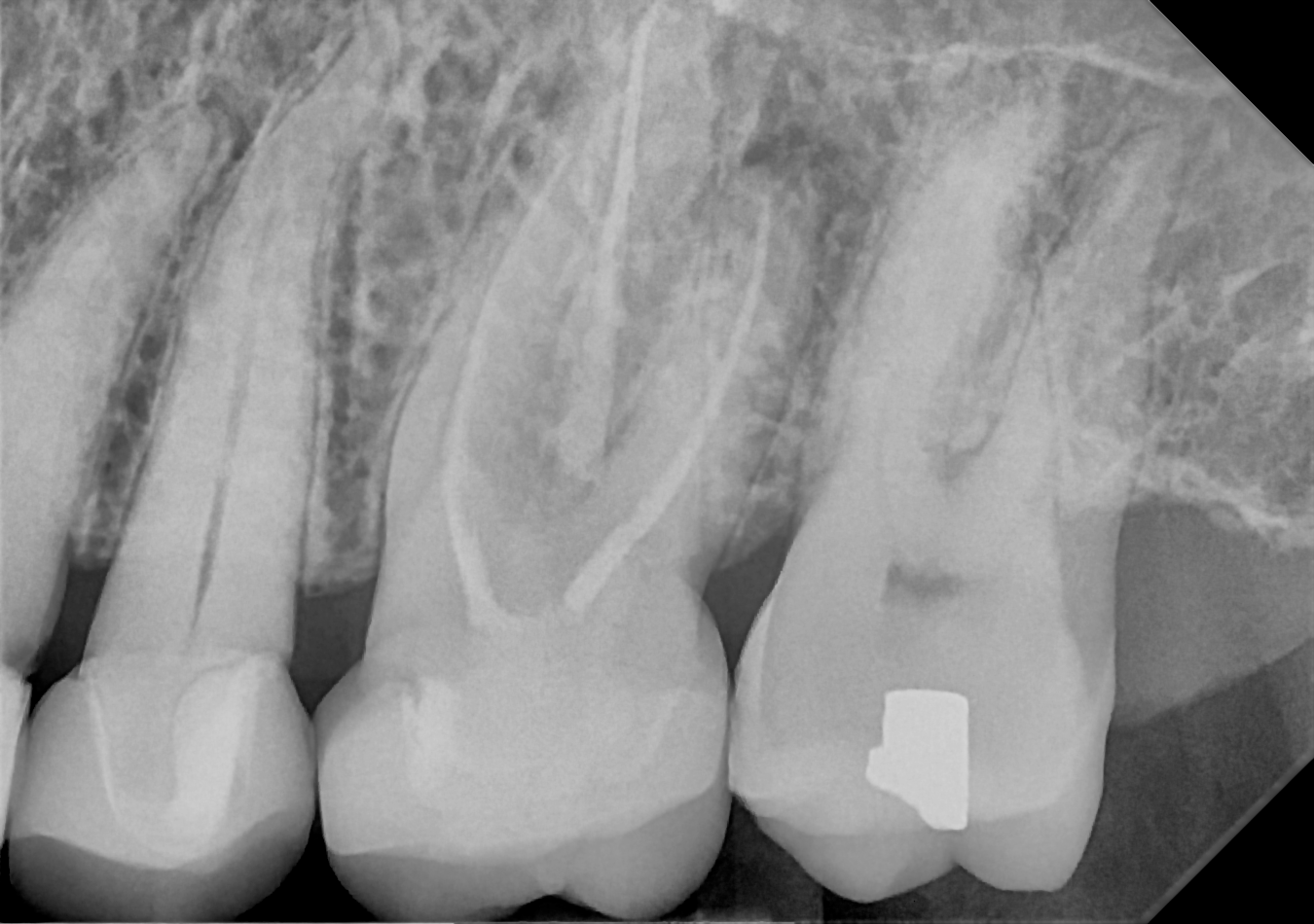
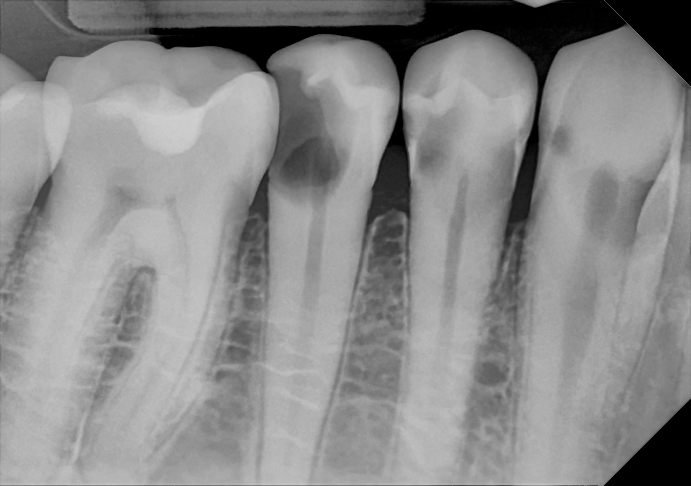
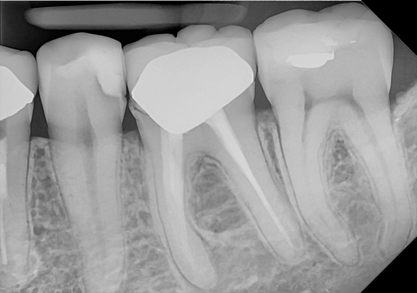
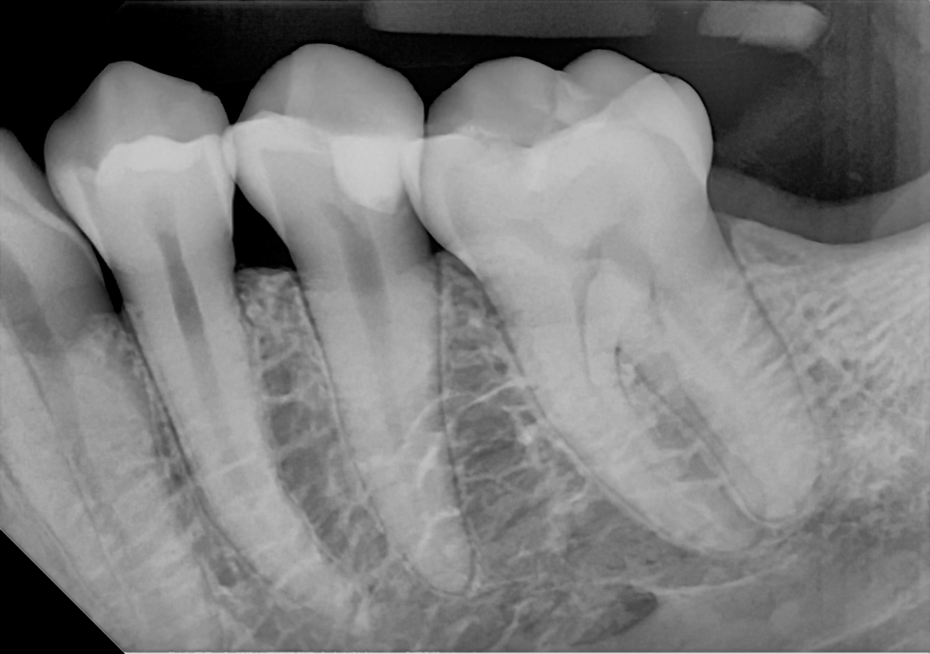
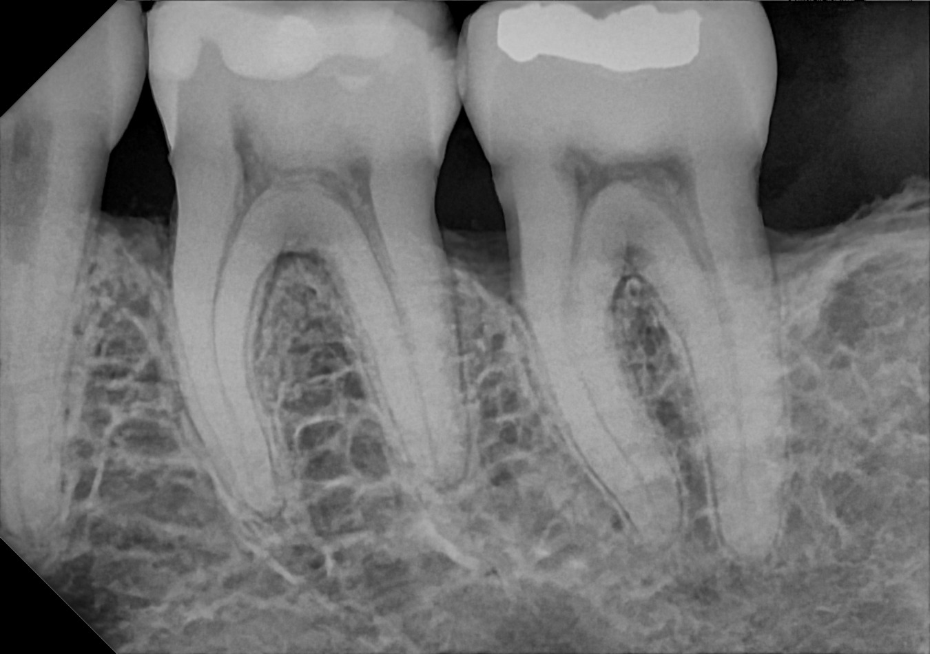
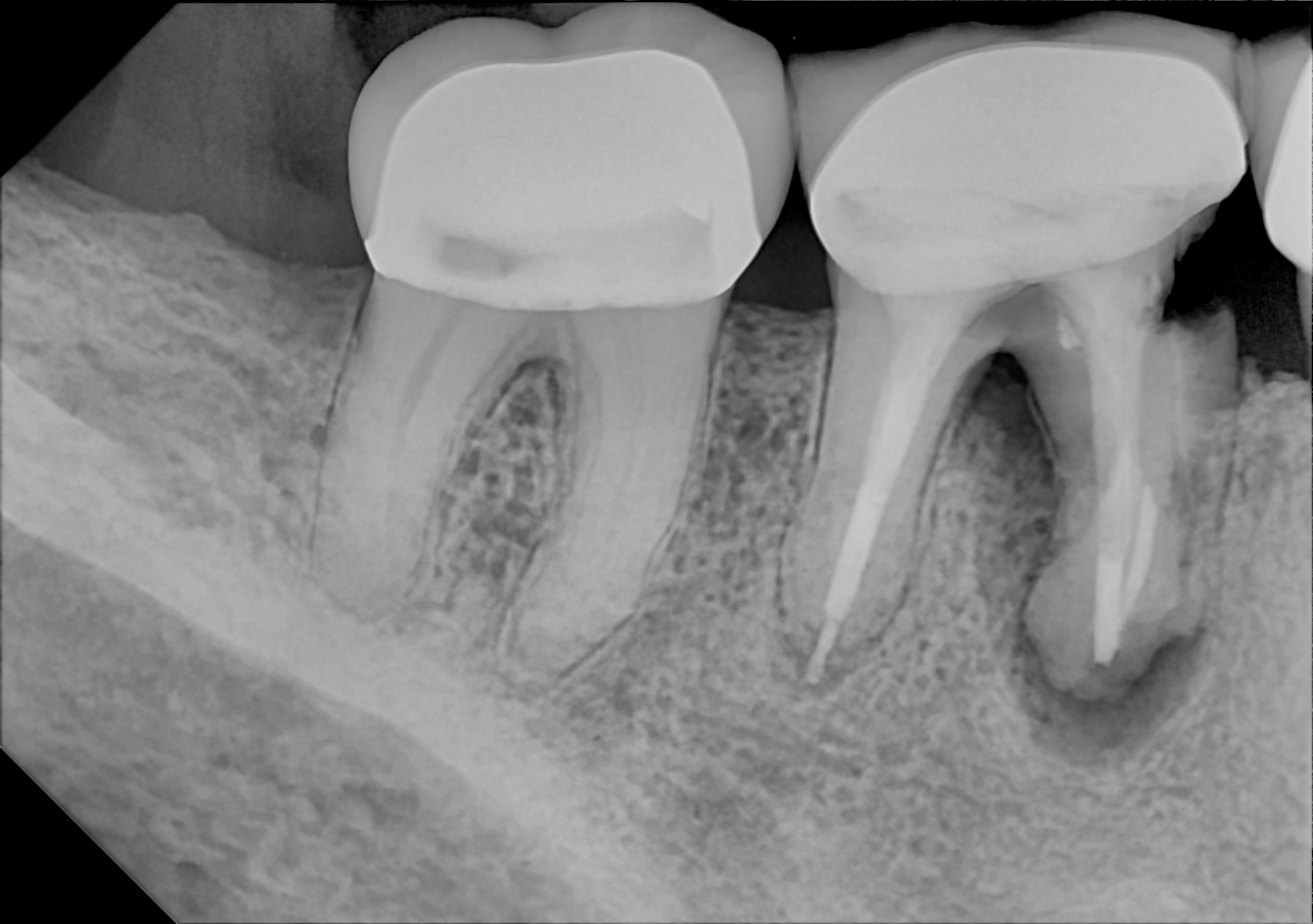
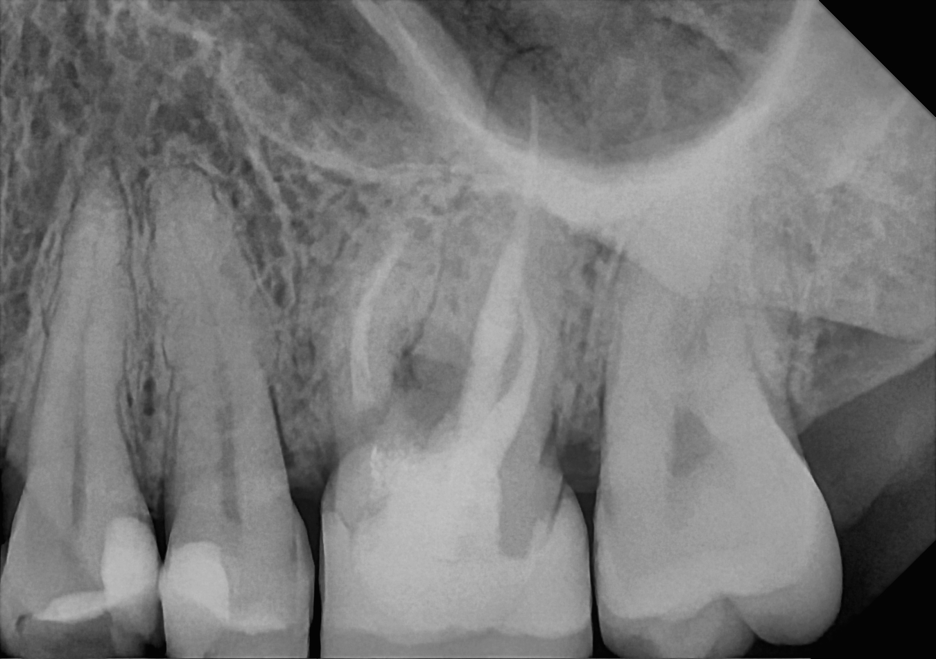
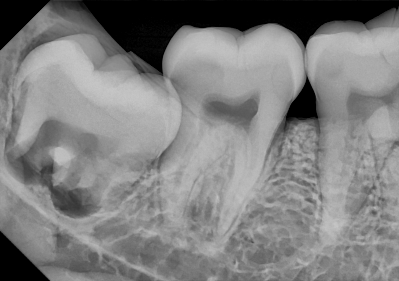
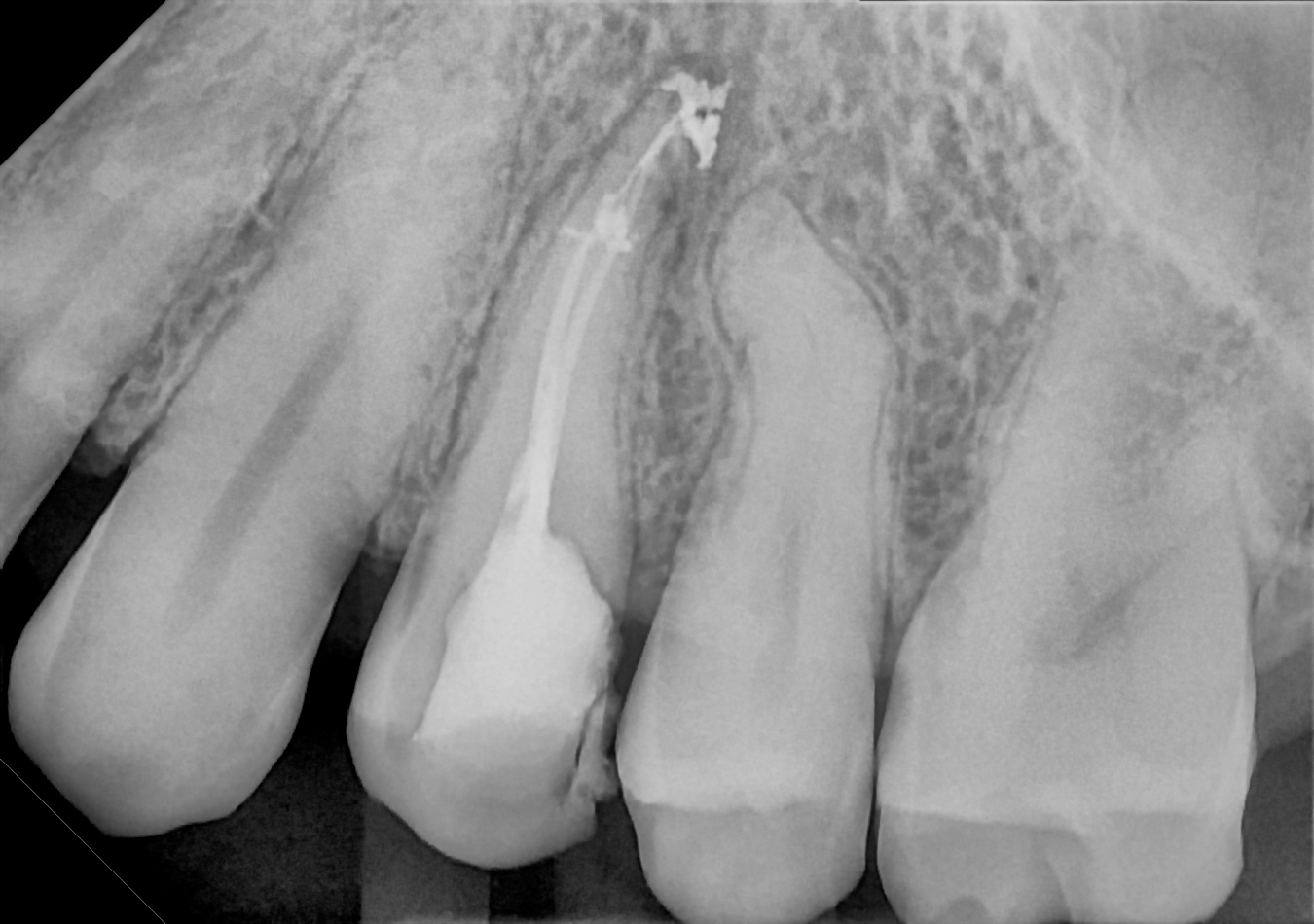
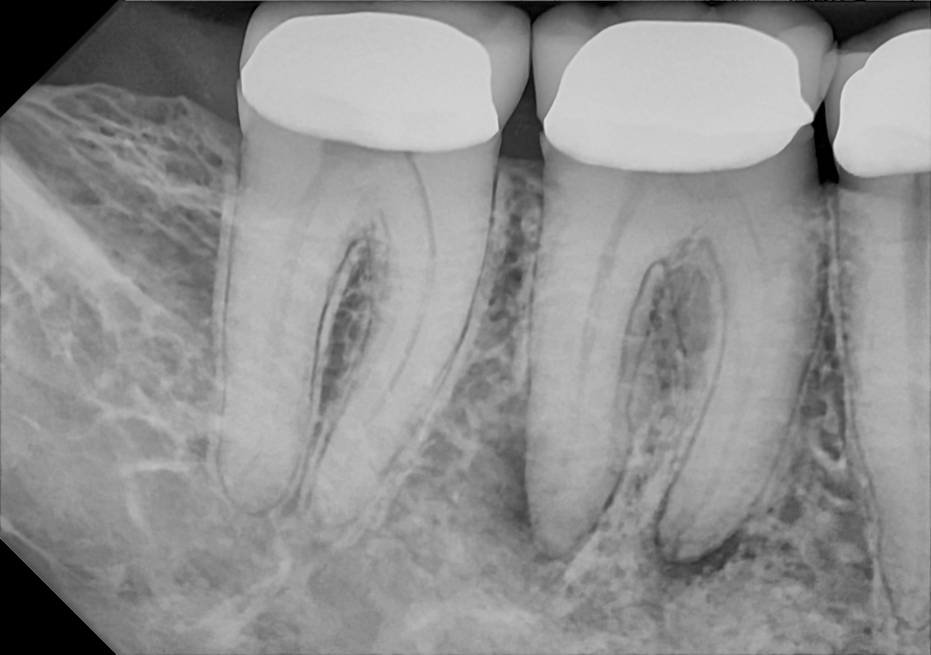
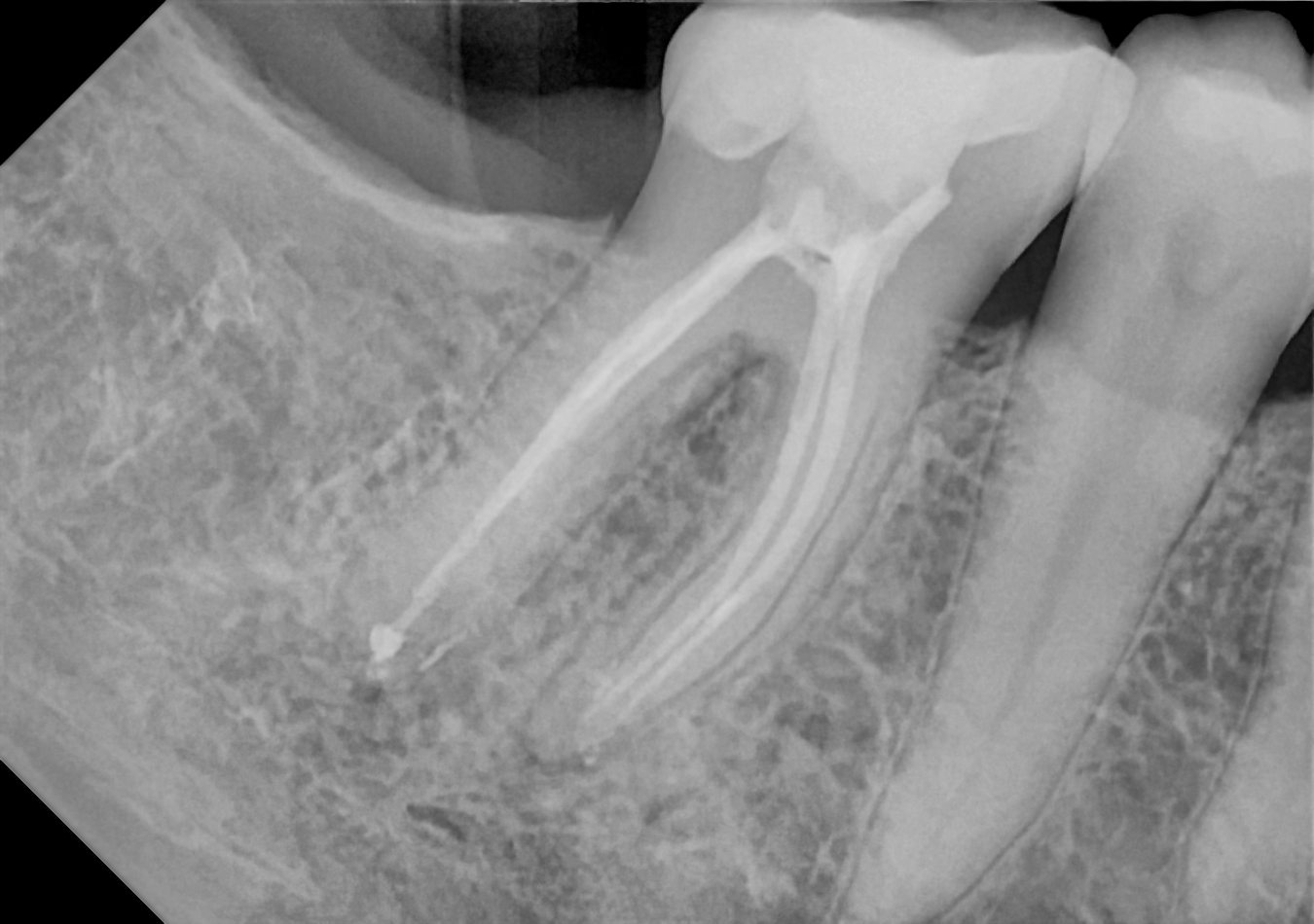
Image gallery
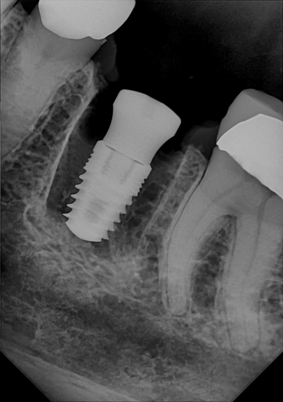
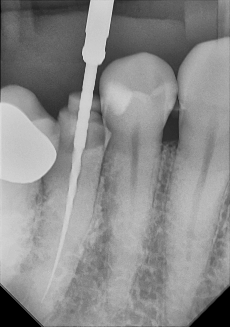
Specialty Shots
The Zero Profile® Endodontic holder makes difficult images easier with additional positioning flexibility and maximum patient comfort.
Let’s get started
Ready to upgrade your imaging?
Try the True Wireless® revolution in your own clinical workflow.